


钨(W)是聚变装置中最具有应用前景的等离子体材料(Plasma facing material,PFM)。因此,研究在极端且复杂的辐照环境中钨与聚变反应产物氦(He)离子间的相互作用有重要的意义。聚变装置运行时,PFM承受高热流(约2~10 MW/m2)和高通量等离子体(1021~1024 m-2s-1)的轰击[1,2],严重影响其热力学性能和机械强度[3,4]。低能氦离子辐照钨材料,在其表面产生氦泡、孔洞和纳米丝等微观结构[5~7]。这些微观结构的形成,与He泡生长、破裂等行为有显著的相关性[8,9]。研究结果表明[10~15],W表面钨丝等微观形貌的演变强烈依赖He离子的入射能量、离子剂量和W样品的温度等参数。W样品的温度低于900 K时,在其表面形成针孔形貌;温度为900~2000 K在W表面生成类似珊瑚结构的纳米丝,称为“钨丝”,且随着温度的升高钨丝层的厚度增加[8,16]。形成钨丝结构的最低离子能量约为20 eV,并且能量的增加使钨丝层的厚度增加,但是高于W溅射阈值的能量会引起表面溅射和侵蚀。给定离子的能量和温度,钨丝层的厚度与离子剂量呈正相关[17,18]。温度高于2000 K时在W样品表面会形成微米尺度肿胀/氦泡/孔洞[5,8,16]。温度是影响W表面形貌的关键因素。已有研究表明,W表面的温度是产生热空位的必要条件[19]。热空位在氦泡生长中起至关重要的作用,样品温度的升高使热空位的密度迅速提高,有利于氦泡成核。但是,在过高的温度(高于2000 K)下空位易迁移而使氦泡在材料表面附近解离,从而抑制氦泡和钨丝的生长[16]。W表面纳米丝的生长温度(T)范围为 0.25 < T/Tm < 0.6,Tm为W的熔点(~3683 K),即920 K < T < 2209 K。对温度低于2000 K时钨表面氦泡和钨丝生长的演变,已有大量的研究[8,12,20,21]。在服役过程中钨材料表面的钨丝结构很容易被轰击而掉到等离子体中,使热核聚变反应的不稳定和不可控。因此,深入了解在钨丝生长温度范围内W中的He泡和W表面钨丝的生长,对于预测在复杂环境下W材料的服役及辐照损伤特性至关重要。本文研究在高温(> 2000 K)条件下氦离子剂量、能量和温度等参数对钨表面形貌的影响,以及W材料的辐照损伤特性。
1 实验方法
实验用钨块的纯度为99.95%,体积为10 mm ×10 mm × 2 mm。在磨抛机上采用十字交错的方式将钨块样品依次用320目、600目、1500目、2000目和5000目砂纸磨平整使其有光泽,然后用粒度为w2.5的金相抛光膏将其表面抛光成镜面,再将样品放入酒精溶液中超声波清洗15~30 min,然后在干燥箱中烘干后冷却至室温得到金相试样,将其密封备用。
离子辐照实验在射频(Radio frequency,RF)驱动的电感耦合等离子体(Inductively coupled plasma,ICP)源放电系统中进行[21,22],其结构示意图如图1所示。用真空泵组维持系统主室的背景压力约为1.0 × 10⁻⁴ Pa,实验时将高纯度(> 99.999%)氦气在5.5 Pa的压力下经气路系统引入离子源,在频率为2 MHz、射频功率为21.4~30 kW的条件下产生氦等离子体。辐照实验所用氦离子的能量范围为30~120 eV,氦离子通量范围为5.1 × 10²²~1.3 × 10²³ m-2s-1,样品的温度范围为2100~2400 K。氦离子的剂量为离子通量与辐照时间的乘积。实验中,采用控制变量法研究辐照参数的影响。研究样品温度的影响时,保持离子的能量和剂量不变。实验结束后关闭工作气体,使样品在真空条件下自然冷却至室温。作为对照,进行2组氢等离子体放电实验(No.18-19),实验参数列于表1。
图1

表1 He离子辐照实验的主要参数
Table 1
| No. | RF power / kW | Ion flux / m-2·s | Ion energy / eV | Ion fluence / m-2 | Irradiation time / s | Surface temperature / K | Gas |
|---|---|---|---|---|---|---|---|
| 1 | 30 | 1.3 × 10²³ | 30 | 4.0 × 1024 | 31 | 2300 | He |
| 2 | 30 | 1.3 × 10²³ | 30 | 1.0 × 1025 | 77 | 2300 | He |
| 3 | 30 | 1.3 × 10²³ | 30 | 3.0 × 1025 | 231 | 2300 | He |
| 4 | 30 | 1.3 × 10²³ | 30 | 5.0 × 1025 | 385 | 2300 | He |
| 5 | 30 | 1.3 × 10²³ | 30 | 1.0 × 1026 | 770 | 2300 | He |
| 6 | 21.4 | 7.4 × 10²² | 80 | 1.0 × 1025 | 120 | 2300 | He |
| 7 | 21.4 | 7.4 × 10²² | 80 | 3.0 × 1025 | 376 | 2300 | He |
| 8 | 21.4 | 7.4 × 10²² | 80 | 5.0 × 1025 | 690 | 2300 | He |
| 9 | 21.4 | 7.4 × 10²² | 80 | 7.0 × 1025 | 952 | 2300 | He |
| 10 | 21.4 | 7.4 × 10²² | 80 | 1.0 × 1026 | 1360 | 2300 | He |
| 11 | 21.4 | 7.4 × 10²² | 80 | 2.0 × 1026 | 2719 | 2300 | He |
| 12 | 20 | 5.1 × 10²² | 80 | 1.0 × 1025 | 210 | 2100 | He |
| 13 | 20.6 | 5.8 × 10²² | 120 | 1.0 × 1025 | 180 | 2300 | He |
| 14 | 29 | 1.2 × 10²³ | 80 | 1.0 × 1025 | 95 | 2400 | He |
| 15 | 29 | 1.2 × 10²³ | 80 | 3.0 × 1025 | 250 | 2400 | He |
| 16 | 29 | 1.2 × 10²³ | 80 | 5.0 × 1025 | 420 | 2400 | He |
| 17 | 29 | 1.2 × 10²³ | 80 | 1.0 × 1026 | 720 | 2400 | He |
| 18 | 30 | 1.3 × 10²³ | 30 | 1.0 × 1026 | 770 | 2300 | H2 |
| 19 | 26 | 7.7 × 10²² | 80 | 1.0 × 1026 | 1278 | 2300 | H2 |
用扫描电子显微镜(型号Hitachi S4800,SEM)表征W样品的表面形貌。样品表面的肿胀/孔洞形貌均是氦泡生长和破裂引起的,因此将肿胀/孔洞的尺寸和数量等参数均作为氦泡的相应参数并加以统计。统计方法:根据SEM照片用Nano Measurer软件的Mark功能,用直线逐一手动标记表面肿胀/氦泡/孔洞得到其半径的统计数据和氦泡的数量,计算氦泡的平均大小和数量密度。用分析天平(MS205DU,精度0.1 mg)测量W样品辐照前后的质量。
2 结果和讨论
2.1 离子剂量对钨样品表面形貌的影响
在离子能量为30 eV、样品温度为2300 K条件下研究了离子剂量对W样品表面形貌的影响。图2不同He离子剂量辐照后W样品表面的SEM照片,图2a给出了辐照前W样品的表面微观结构。从SEM图像中可见,W样品表面有一些宽度为几十纳米的直线,是机械抛光造成的划痕。离子剂量为4.0 × 1024 /m2时,在W样品表面出现大小不同的表面肿胀(半径为20~70 nm) (图2b),表明在放电过程中随着氦原子在W基体中的积累氦泡开始发展并在表面形成肿胀。离子剂量增加到1.0×1025 /m2,氦泡趋于在表面均匀分布(图2c),其尺寸和密度明显增加,平均半径约为106 nm。随着离子剂量从1.0 × 1025 /m2 提高到5.0×1025 /m2 (图2d,e),氦泡迅速增大其密度随之提高。离子剂量增加到5.0 × 1025 /m2,使氦泡的平均半径增加到约155 nm。值得注意的是,大部分氦泡是独立生长的,但是相邻的氦泡相互间的距离越来越小。离子剂量增加到1.0 ×1026 /m2 (图2f),在氦泡的表面出现孔洞,部分氦泡相互接触产生类似珊瑚状交联的纳米丝结构[8,23,24],但是纳米丝的尺寸较大,是钨丝的初始生长阶段。
图2

图2
不同 He 离子剂量辐照后W样品表面的 SEM 照片
Fig.2
SEM analysis of the surface morphology of W samples irradiated by He ion with different fluence (a) 0 /m2, (b) 4.0 × 1024 /m2, (c) 1.0 × 1025 /m2, (d) 3.0 × 1025 /m2, (e) 5.0 × 1025 /m2 , (f) 1.0 × 1026 /m2
图3

图3
离子剂量为4.0 × 1024 /m2时W样品表面氦泡的半径分布、对直方图的高斯拟合结果以及氦泡平均半径和数量密度与离子剂量的关系
Fig.3
Histogram shows the radius distribution of He bubbles in the W sample surface at a fluence of 4.0 × 1024 /m2, the solid line shows the Gaussian fitting results of the histogram (a), and relationship between the average radius and density of He bubbles and ion fluence (b, c)
以上实验结果表明,在样品温度为2300 K的条件下,高温氦离子辐照后在W的表面出现大小约为40~150 nm的肿胀/孔洞,继续提高离子剂量则肿胀/孔洞演变成较粗的初始钨丝状结构。用离子辐照钨材料,先是氦原子注入到材料中,热扩散使氦原子到达样品体相中更深的位置。氦原子被材料中的缺陷捕获形成一定尺寸的氦泡,在表面产生肿胀的形貌并伴随产生自间隙钨原子。随着氦泡的生长氦泡内的压力提高而挤压氦泡周围的材料,压力超过钨材料的断裂阈值时氦泡破裂而形成孔洞,使材料表面越来越粗糙。高密度氦泡不断在钨用品的近表面生长和破裂,最终在表面形成珊瑚状交联的纳米丝状结构。
2.2 离子能量对钨样品表面形貌的影响
在离子剂量为1.0 × 1025 /m2、样品温度为2300 K的条件下,研究了氦离子的能量对W样品的表面形貌的影响。氦离子对钨材料的溅射阈值能为约120 eV[12],因此实验中三种离子的能量分别为30、80和120 eV,图4给出了不同能量的氦离子辐照后W样品的表面SEM照片。可以看出,氦离子能量为30 eV时,图4a中W样品的表面出现了氦泡,其生长产生了纳米肿胀/孔洞(半径为50~100 nm)。随着离子能量增加到80 eV (图4b),W样品表面出现了交联的起伏形貌并形成了明显的孔洞。产生孔洞的原因是,大量氦原子被捕获聚集形成氦泡,然后在材料的表层破裂。随着离子能量增加到120 eV,W样品表面形成了初始的钨纳米丝结构,大部分孔洞被钨纳米丝状结构遮住(图4c)。实验结果表明,随着离子能量的增加氦离子注入表层的深度增加,最终停留在W样品的近表层促进了表层氦泡的生长、破裂及肿胀,其结果是在W样品表面产生纳米丝状结构。
图4

图4
不同 He 离子能量辐照后W样品表面的SEM照片
Fig.4
SEM analysis of the surface morphology of W samples irradiated by He ion with different ion energy (a) 30 eV, (b) 80 eV, (c) 120 eV
研究了样品温度为2300 K、离子能量为80 eV的条件下,离子剂量的变化对W样品的表面形貌的影响。图5给出了氦离子辐照后W样品的表面SEM照片,可以看出,离子剂量大于1.0 × 1025 /m2时在W样品表面均产生了明显的钨丝状结构。氦离子剂量为1.0 × 1025 /m2 时(图 5a),W样品表面的钨丝状结构开始发展。随着离子剂量从3.0×1025 /m2增加到5.0 × 1025 /m2 (图5b,c),W样品表面的钨纳米丝逐渐致密,随着离子剂量从7.0 × 1025 /m2增大到2.0 ×1026 /m2 (图5d~f),钨丝状结构的密度逐渐稳定。钨丝状结构的生长模式表明,在低剂量(< 5.0 × 1025 /m2)条件下钨丝状结构的生长速率较高,超过这个剂量后钨丝状结构的生长速率逐渐降低。其原因是,钨层达到一定厚度后密集的钨丝状结构可能使进入钨丝状结构底部到达样品近表面层的He离子的数量和能量减小,从而使钨丝状结构的生长速率下降。
图5

图5
不同He离子剂量辐照后W样品表面的SEM照片
Fig.5
SEM analysis of the surface morphology of W samples irradiated by He ion with different fluence (a) 1.0 × 1025 /m2, (b) 3.0 × 1025 /m2, (c) 5.0 × 1025 /m2, (d) 7.0 × 1025 /m2, (e) 1.0 × 1026 /m2, (f) 2.0 × 1026 /m2
2.3 样品的温度对钨样品表面形貌的影响
在离子能量为80 eV、离子剂量为1.0 × 1025 /m2的条件下,研究了样品温度对W样品的表面形貌的影响。图6给出了辐照后W样品的表面SEM照片,图6a、b中样品的温度为2100 K,图6b是在样品倾斜一定角度后拍摄(与水平方向夹角~30°)的,图6c、d中样品的温度分别为2300和2400 K。样品温度为2100 K时在W样品表面观察到明显交叉关联的珊瑚状的纳米丝(图6a、b),温度提高到2300 K时(图6c)表面钨丝状结构处于开始发展的状态。继续提高温度到2400 K (图6d),在表面只能看到氦泡破裂后产生的孔洞。随着样品温度从2100 K升高到2400 K,表面氦泡/钨丝状结构的密度明显降低。在辐照期间W样品表面的氦泡生长的同时产生了大量自间隙钨原子,其在高温下极不稳定。实验结果表明,当温度高于2000 K、离子剂量相同时,材料表面温度的提高加强了表面自间隙钨原子在材料表层的融合和扩散,从而抑制了表面肿胀和钨丝状结构生长,甚至使钨丝状结构退化。同时,过高的温度可能使部分氦泡解离和氦泡/钨丝状结构的密度下降[17]。
图6
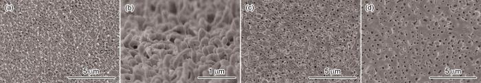
图6
不同温度He离子辐照后W样品表面的SEM照片
Fig.6
SEM analysis of the surface morphology of W samples irradiated by He ion with different temperature (a, b) 2100 K, (c) 2300 K (d) 2400 K
图7

图7
不同He离子剂量辐照后W样品表面的SEM照片
Fig.7
SEM of the surface morphology of W samples irradiated by He ion with different fluence (a) 1.0 × 1025 /m2, (b) 3.0 × 1025 /m2, (c) 5.0 × 1025 /m2, (d~f) 1.0 × 1026 /m2
图8

图8
氢离子辐照后W样品表面的SEM照片
Fig.8
SEM analysis of the surface morphology of W samples irradiated by hydrogen ion (a) H2-30 eV-2300 K, (b) H2-80 eV-2300 K
实验结果表明,高温氦离子辐照后,钨材料中氦泡的生长和破裂使表面形貌发生变化。同时,钨丝状结构的产生和演变与氦泡的形成、生长及破裂有密切的关系。
2.4 辐照引起的钨样品的质量损失
在不同辐照参数下W样品的质量损失为
式中m1和m2分别为辐照前后W样品的质量,S为辐照有效面积。根据质量损失可计算出溅射率(Sputtering yield,YS)
式中MA为W的原子质量,t为辐照时间,Г为离子通量。
图9给出了辐照后W样品质量损失的计算结果。图9a表明,在离子能量为30 eV、样品温度为2300 K条件下质量损失几乎不随离子剂量变化。但是,在离子能量为80 eV、温度为2300和2400 K条件下W样品的质量损失都随着He离子剂量的增加而增加,并且质量损失几乎是线性增加。在实验的离子剂量范围内,温度为2400 K时的质量损失均比温度为2300 K时的高。在离子能量为80 eV、离子剂量为1.0 × 1025 /m2条件下样品温度为2300和2400 K时的质量损失分别为1.69和2.08 g/m2 (图9b),而随着离子剂量的增至1.0 × 1026 /m2温度为 2400 K的质量损失(8.77 g/m2)是2300 K时(4.38 g/m2)的约2倍。
图9

图9
W样品的质量损失与He离子剂量和He离子能量的关系
Fig.9
Mass loss of W samples as a function of He ion fluence (a) and He ion energy (b)
高能He离子与W表面原子的高频碰撞使W表层原子发生溅射,且随着He离子辐照剂量的增加溅射从表层向下延伸。但是,珊瑚状交联的纳米丝状结构较大的比表面积能有效屏蔽He离子的注入并具有极高的抗溅射能力[34,35]。这种结构可避免高能 He侵蚀W的基底,对W基底有一定的保护作用。质量损失主要是离子能量轰击产生的溅射引起的,因此也影响样品的质量损失。由图5可见,当样品温度为2300 K、离子能量为80 eV时,W样品表面产生更明显的钨丝状结构结构,W纳米丝交联网状结构能在一定程度上避免高能He离子侵蚀W样品的基底。但是,在样品温度为2400 K、离子能量为80 eV的条件下(图7a~d)表面W丝与W丝之间交联的网状结构较少,对W样品基底的保护明显减弱。同时,温度的升高也提高了样品表面最外层自间隙钨原子的瞬间蒸发速率,从而使2400 K时样品的质量损失比2300 K时的高。
在样品温度为2300 K、离子剂量为1.0 × 1025 /m2的条件下,随着离子能量从30 eV提高到120 eV质量损失从1.54 g/m2增加到2.67 g/m2 (图9b)。表2 给出了不同能量的He离子辐照后W样品溅射率变化。可以看出,随着离子能量的增加W表面的溅射率也随之提高,使质量损失更加显著。质量损失的计算结果表明,在高温氦离子辐照过程中,氦泡的生长和表面钨丝状结构的产生使W样品表面发生了明显的侵蚀。在高温辐照期间氦泡生长挤出的自间隙钨原子在表面极不稳定,原子之间远低于正常体相中钨原子的结合能使W表面的溅射阈值低于体相内钨原子。因此,受到低能氦离子的轰击也产生明显的溅射,并且愈接近溅射阈值的能量产生的溅射越明显,使质量损失随溅射的变化产生了相似的演变趋势。
表2 不同 He 离子能量辐照后 W 样品的溅射率
Table 2
| Ion energy / eV | Ion fluence / m-2 | Mass loss / g·m-2 | Ys |
|---|---|---|---|
| 30 | 1.0 × 1025 /m2 | 1.54 | 5.0 × 10⁻⁴ |
| 80 | 1.0 × 1025 /m2 | 1.69 | 6.2 × 10⁻⁴ |
| 120 | 1.0 × 1025 /m2 | 2.67 | 8.4 × 10⁻⁴ |
3 结论
(1) 在2100~2400 K温度区间低剂量氦离子辐照后钨样品表面产生氦泡/肿胀形貌和氦泡破裂形成的孔洞,随着离子剂量的提高逐渐形成类似珊瑚状交联的纳米丝状结构。
(2) 高温促进表面自间隙钨原子的扩散而抑制钨丝状结构的生长。在温度(2300 K)和离子剂量(1.0 × 1025 /m2)相同的条件下,氦离子能量从30 eV增加到120 eV注入深度随之增加并促进表面氦泡和钨丝状结构的生长。
(3) 离子能量的增加使样品表面的溅射率提高,从而使质量损失增加,表面交联的钨丝状结构可减少质量损失。在高温氦离子辐照下,钨形貌的演变主要受温度、离子能量和剂量的影响。
参考文献
Progress of the European R&D on plasma-wall interactions, neutron effects and tritium removal in ITER plasma facing materials
[J].
Material erosion at the vessel walls of future fusion devices
[J].
Physics basis and design of the ITER plasma-facing components
[J].
Plasma operation with tungsten tiles at the central column of ASDEX Upgrade
[J].
Helium effects on tungsten under fusion-relevant plasma loading conditions
[J].
Effect of tungsten crystallographic orientation on He-ion-induced surface morphology changes
[J].
He-ion and self-atom induced damage and surface-morphology changes of a hot W target
[J].
Helium effects on tungsten surface morphology and deuterium retention
[J].
Helium concentration measurement in tungsten fuzz-like nanostructures by means of thermal desorption spectroscopy
[J].
Formation process of tungsten nanostructure by the exposure to helium plasma under fusion relevant plasma conditions
[J].
TEM observation of the growth process of helium nanobubbles on tungsten: nanostructure formation mechanism
[J].
Helium induced nanoscopic morphology on tungsten under fusion relevant plasma conditions
[J].
Nanostructure formation on tungsten exposed to low-pressure rf helium plasmas: a study of ion energy threshold and early stage growth
[J].
Nanostructuring of molybdenum and tungsten surfaces by low-energy helium ions
[J].
Comparison of tungsten nano-tendrils grown in Alcator C-Mod and linear plasma devices
[J].
Helium, hydrogen, and fuzz in plasma-facing materials
[J].
Research on tungsten material irradiation damage induced by low-energy and high-flux hydrogen/helium ions
[D].
低能强流氢氦离子辐照下钨材料损伤研究
[D].
Impact of helium ion energy modulation on tungsten surface morphology and nano-tendril growth
[J].
Formation mechanism of bubbles and holes on tungsten surface with low-energy and high-flux helium plasma irradiation in NAGDIS-II
[J].
Helium and deuterium implantation in tungsten at elevated temperatures
[J].
High-flux He+ irradiation effects on surface damages of tungsten under ITER relevant conditions
[J].
Stress-driven surface swell and exfoliation of copper as the plasma-facing materials in NBI ICP source
[J].
W nano-fuzz growth by high-flux He ion irradiation with their energy above 300 eV
[J].
Effect of temperature on the growth and surface bursting of He nano-bubbles in W under fusion-relevant He ion irradiations
[J].
Investigation of hydrogen bubbles behavior in tungsten by high-flux hydrogen implantation
[J].
In-situ TEM observation of the evolution of helium bubbles in Mo during He+ irradiation and post-irradiation annealing
[J].
The effect of displacement damage on deuterium retention in ITER-grade tungsten exposed to low-energy, high-flux pure and helium-seeded deuterium plasmas
[J].
The effect of ion energy and substrate temperature on deuterium trapping in tungsten
[D].
Research on micro-nano scale damage on tungsten surface under helium irradiation
[D].
氦辐照条件下钨材料表面微纳尺度的损伤研究
[D].
Near-surface thermal characterization of plasma facing components using the 3-omega method
[J].
Effects of sequential tungsten and helium ion implantation on nano-indentation hardness of tungsten
[J].
Thermal conductivity reduction of tungsten plasma facing material due to helium plasma irradiation in PISCES using the improved 3-omega method
[J].
Effects of helium on mechanical properties of tungsten for fusion applications
[J].
Effect of helium ions irradiation on stability of nano-tungsten whiskers
[J].
Nanoscale tungsten whiskers grown on the surface of polycrystalline W-plate was subjected intermittently to irradiation of He ions with energy of 150 eV at 400 K. The effect of He ion irradiation on the evolution of nanoscale W-whiskers were investigated by means of scanning electron microscope, transmission electron microscope and mass loss method. The results show that nanoscale W-whiskers were extremely unstable in the course of high-energy He ion irradiation, and the degree of crosslinking between W-whiskers decreases gradually with the increase of irradiation fluence. Due to high-energy He ion sputtering, He bubbles existed in the whiskers will break and lead to collapse and coalesce of the W-whiskers. Meanwhile, certain amount of the yielded W atoms by He ion sputtering may re-deposited on the outer wall or the root of the nanoscale whiskers nearby, and finally, the relevant nanoscale whiskers may evolve into a cone-shaped structure with a thin top and a thick root.
氦离子辐照对钨纳米丝稳定性的影响
[J].用150 eV高能氦(He)离子在400 K对多晶钨(W)表面的W纳米丝进行间歇式辐照并使用扫描电子显微镜、透射电子显微镜以及称重法等手段对其表征,研究了He离子辐照对W纳米丝演变过程的影响。结果表明,高能He离子辐照使W纳米丝极不稳定。随着辐照剂量的增加W纳米丝之间的交联程度逐渐降低。W丝内的He泡在高能He离子溅射的作用下破裂,使W丝塌陷合并,部分溅射出来的W原子沉积在近邻的W纳米丝外壁或W丝根部,最终使W纳米丝演变成顶部细根部粗的锥型结构。
W fuzz layers: very high resistance to sputtering under fusion-relevant He+ irradiations
[J].
