


琼脂糖(AG)是从海洋红藻中提取出来的多糖,具有生物相容性,含有重复1,3连结的β-D-半乳糖和1,4连结的3,6-内醚-L-半乳糖。使用琼脂糖可制备热可逆凝胶[3,4]。由于氢键的作用,不使用京尼平这类有毒交联剂即可生成琼脂糖水凝胶[5],使其成为一种生物相容性聚合物[6]。因此,用琼脂糖及其衍生物和混合物制备的水凝胶有望用于组织工程和再生医学[7],如神经发生[8],软骨形成,骨再生[9],伤口愈合[10]。基于琼脂糖的生物水凝胶,对特定组织工程应用具有多功能性能[11]。但是单一琼脂糖构成的水凝胶机械性能差,不能满足实际需要。为了制备具有良好机械性能的琼脂糖水凝胶,可将琼脂糖与其它聚合物共混制成复合型水凝胶以改善其性能。Brandon等[12]将琼脂糖和聚乙二醇双丙烯酸酯结合生成一种互穿网络水凝胶,其剪切模量比纯琼脂糖网络增大了4.9倍(8.2 kPa);Kun Yan等[13]将壳聚糖掺入琼脂糖基质中制备出具有高度多孔结构的新型互穿聚合物网络水凝胶,其孔隙率高于95%,水诱导形状恢复率高于90%。
1 实验方法
1.1 实验用试剂和仪器
实验用仪器有,傅里叶漫反射红外光谱仪(FT-IRPrestige-21);冷冻干燥仪(VFD-21S);扫描电镜(SEM)(JSM6510LV);X射线衍射仪(XRD)(AL-Y3000)。
1.2 AG/PVA/CB[7]水凝胶的制备
AG和PVA水凝胶的制备:将0.3 g AG放入50 mL烧杯后加入5 mL去离子水,加热使其充分溶解后倒入模具,冷却至室温后得到AG水凝胶。将0.3 g PVA和5 mL蒸馏水加入烧杯后加热,PVA充分溶解后倒入模具,将其在-20 ℃冷冻14 h,取出后在室温解冻1 h,此为一个冷冻-解冻循环过程。制备不同冷冻-解冻次数的传统PVA水凝胶样品。
AG/PVA/CB[7]的制备:取5 mL去离子水和0.3 g AG,将0.3 g PVA和0.02 g CB[7]放入50 mL烧杯中,超声震荡5 min使CB[7]均匀分散于溶液中,密封后进行90℃水浴加热搅拌,待其完全溶解后置入模具中。使其冷却至室温后放入-20℃冷冻箱中冷冻14 h成型,然后在室温下解冻1 h,此为一个冷冻-解冻循环过程。改变样品的组成和冷冻-解冻次数,制备相应的 AG/PVA/CB[7]水凝胶样品。
1.3 AG/PVA/CB[7]水凝胶的SEM、FT-IR和XRD表征
将冷冻干燥所得的干凝胶放入液氮中焠断,得待测凝胶样品。对其表面喷金120 s,用扫描电镜观察样品表面的形貌结构;分别取适量样品和KBr压片,进行红外光谱(FT-IR)测试;将水凝胶真空干燥后磨碎测试XRD谱。使用XRD衍射仪在40 kV和200 mA条件下产生的CuKα,衍射角(2θ)为5°~90°,扫描速度为2°/min,检测样品的结晶情况。
1.4 AG/PVA/CB[7]水凝胶的力学性能测试
使用模具制成2 mm厚的样品,将其切割使其尺寸为12 mm×2 mm×2 mm。使用WDW-BO5型万能试验机进行拉伸测试,拉伸速率为1 mm·s-1,应力和应变为
其中σ为应力,ε为应变,p为压力,A为受力面积,L为拉伸长度,L0为凝胶原始长度。
根据橡胶的熵弹性理论 [19]
计算水凝胶样品的交联密度。式中
1.5 AG/PVA/CB[7]水凝胶自愈性能的测试
将两块长度为5 cm的条状水凝胶样品切成两半,其中一块用罗丹明B染成红色,用滤纸擦去表面的水分,使两块水凝胶的断面紧密接触后放置24 h,对连接在一起的水凝胶进行承重实验,其修复率为
式中δ1,δ2分别为水凝胶自修复前后的断裂前后承重质量。
1.6 AG/PVA/CB[7]水凝胶溶胀性的测试
将干燥后的水凝胶材料称重(W0)后在水中浸泡,并分别在2 h,4 h,6 h,10 h,24 h,28 h,30 h取出,擦去表面水分后称其质量(Wt),反复进行上述操作直到凝胶的质量不变,此为达到了溶胀平衡。水凝胶的溶胀率(SR)为
式中Wt为某时刻水凝胶的质量(g);W0为干燥后的水凝胶的质量(g)。
用上述方法将干燥后的水凝胶材料分别浸于pH=7、不同温度(T=25 ℃,35 ℃,50 ℃,65 ℃)的溶液中,使其达到溶胀平衡。计算水凝胶在不同温度下的溶胀率。
2 结果和讨论
2.1 AG/PVA/CB[7]水凝胶的结构
2.1.1 AG/PVA/CB[7]水凝胶的SEM分析
图1
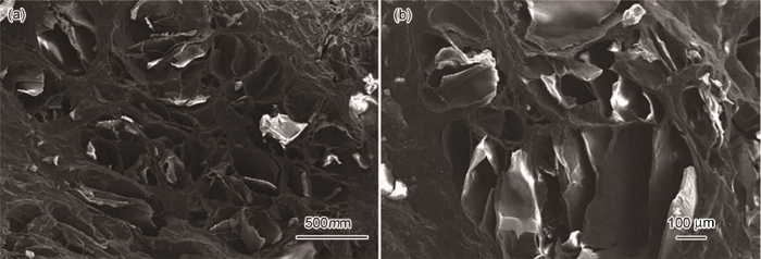
图1
AG/PVA/CB[7]水凝胶的SEM照片
Fig.1
Scanning electron microscopy of AG/PVA/CB[7] hydrogel
2.1.2 红外光谱
图2
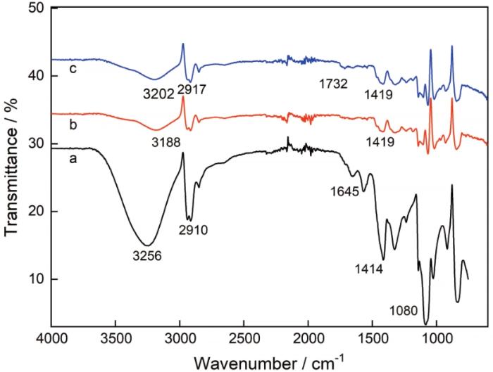
图2
PVA、AG/PVA和AG/PVA/CB[7]水凝胶的红外光谱图
Fig.2
FTIR spectrum of PVA (a), AG/PVA (b), and AG/PVA /CB[7] (c)
2.1.3 AG/PVA/CB[7]水凝胶的XRD谱
图3

图3
AG/PVA/CB[7]水凝胶的XRD谱
Fig.3
X-ray diffraction pattern of AG/PVA/CB[7] hydrogel
图4

2.2 AG/PVA/CB[7]水凝胶的力学性能
2.2.1 组分对AG/PVA/CB[7]水凝胶力学性能的影响
图5给出了不同组分水凝胶的应力-应变曲线。传统AG水凝胶因浓度的限制含水量过大,其结构疏松且脆而易碎,不能用万能试验机测其应力应变曲线。图5表明,PVA/AG水凝胶的力学性能与纯的PVA水凝胶相比有较大的提高,其拉伸应力达到0.32 MPa,比PVA水凝胶提高了39%。其原因是,PVA/AG水凝胶中PVA和AG两个高分子链通过部分氢键和PVA微晶作用形成较高交联密度(交联密度为6.4×10-4 mol·cm-3)的双网络水凝胶结构,其中AG的高分子链为刚性链,PVA链为柔性链。当凝胶受力时刚性网络发生断裂,使能量在凝胶网络中耗散从而保护主体柔性网络不被破坏,维持了凝胶的应力和应变能[21]。同时,图5也表明,CB[7]组分的加入提高了AG/PVA水凝胶的机械性能。其中AG/PVA/CB[7]水凝胶的拉伸强度为0.37 MPa,杨氏模量为 0.23 MPa,均高于AG/PVA水凝胶(其拉伸强度0.32 MPa,杨氏模量0.20 MPa)。其主要原因是,瓜环的端口羰基和两个高分子链通过氢键交联,使PVA和AG高分子链形成更致密的双网络结构。
图5

图5
PVA、AG/PVA和AG/PVA/CB[7]水凝胶的应力-应变曲线
Fig.5
Stress-Strain curves of PVA、AG/PVA and PVA/AG/CB[7] hydrogel
2.2.2 冷冻-解冻次数对AG/PVA/CB[7]水凝胶的力学性能的影响
图6给出了不同冷冻-解冻次数的AG/PVA/CB[7]水凝胶样品的应力-应变曲线。根据图6中的应力-应变曲线可计算出相应样品的交联密度,结果列于表1。从图6可见,随着冻融循环次数的增加应力先增大后趋于平稳。其原因是,复合水凝胶内部的结晶区高分子链有规律排列且分子间距较小,产生较强烈的作用力;而无定形区内的分子链无规律排列。随着冷冻-解冻次数增加样品内的交联点随之增加,交联密度提高,出现更多的结晶微区,使相应的结晶结构更加完整。当冷冻-解冻进行第5次时相应的交联密度最大,微晶区更多,其拉伸应力达到最大值。但是PVA、AG的含量有限,当冻融循环次数增加到使物理交联密度及微晶区数量达到最大值时,拉伸应力趋于平稳。
图6
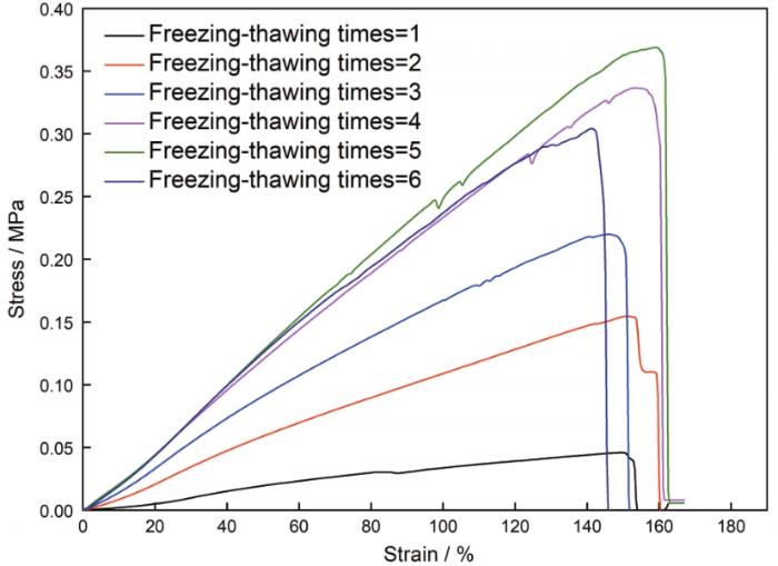
图6
不同冷冻-解冻次数AG/PVA/CB[7]水凝胶力的应力-应变曲线
Fig.6
Stress-strain curves of AG/PVA/CB[7] hydrogel with different freezing-thawing times
表1 AG/PVA/CB[7]水凝胶的交联密度
Table 1
| Freezing-tdawing times | 1 | 2 | 3 | 4 | 5 | 6 |
| Crosslink density /×10-4 mol·cm-3 | 0.93 | 2.9 | 4.5 | 6.6 | 7.2 | 6.4 |
2.3 AG/PVA/CB[7]水凝胶的自愈性能
图7

图8

2.4 AG/PVA/CB[7]水凝胶的溶胀性能
图9
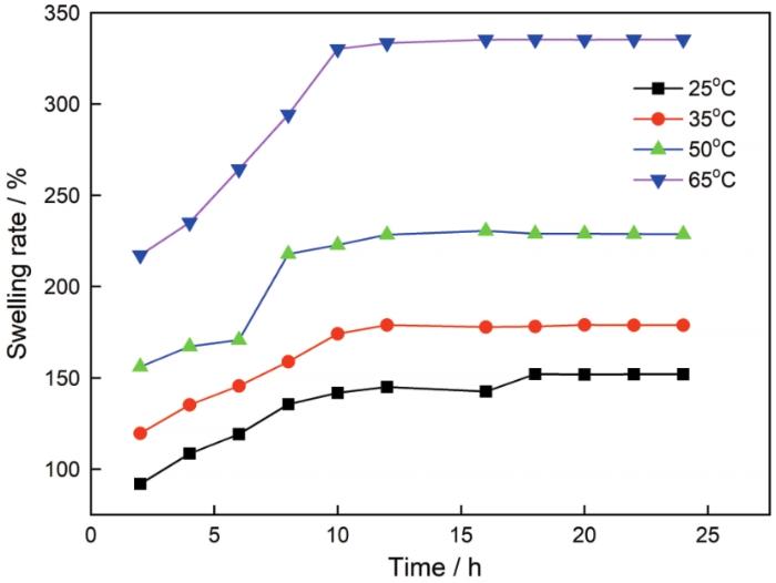
图9
温度对AG/PVA/CB[7]水凝胶溶胀性能的影响
Fig.9
Effects of temperature on the swelling properties of AG/PVA/CB[7] hydrogel
AG/PVA/CB[7]水凝胶中的CB[7]有亲水外壁、PVA和AG链上有羟基等亲水基团。这些亲水基团与越来越多的水溶剂分子以氢键缔合,使水凝胶逐渐膨胀达到平衡状态。随着温度的升高AG/PVA/CB[7]水凝胶中部分氢键作用减弱,聚合物柔性直链舒展,在一定程度上扩大了凝胶网络结构中的孔隙,使更多的水分子进入凝胶网络孔隙中进而提高了溶胀率。
3 结论
使用AG为第一物理交联网络、PVA为第二物理交联网络、CB[7]为交联剂,采用微晶和氢键共同增强策略可制备具有优异力学性能的自愈双网络AG/PVA/CB[7]水凝胶。这是一种强度高、溶胀低且生物相容性好的水凝胶,其力学性能随着冷冻-解冻次数的增加而提高,循环5次后其拉伸强度高达0.37 MPa,其25℃溶胀率只有140%。
参考文献
Fabrication and characterization of electrospun poly-L-lactide/gelatin graded tubular scaffolds: Toward a new design for performance enhancement in vascular tissue engineering
[J].
Controlled delivery of icariin on small intestine submucosa for bone tissue engineering
[J].
Global metabolite profiling of agarose degradation by saccharophagus degradans
[J].
Effect of the hydration on the biomechanical properties in a fibrin-agarose tissue-like model
[J].
How can genipin assist gelatin/carbohydrate chitosan scaffolds to act as replacements of load-bearing soft tissues
[J].
Optimizing structural and mechanical properties of cryogel scaffolds for use in prostate cancer cell culturing
[J].
Hydrogels for skeletal muscle regeneration
[J].
Dental pulp stem cells: advances to applications
[J].
Bioinspired, biomimetic, double enzymatic mineralization of hydrogels for bone regeneration with calcium carbonate
[J].
Agarose bioplastic based drug delivery system for surgical and wound dressings
[J].
We have developed an agarose-based biocompatible drug delivery vehicle. The vehicle is in the form of thin, transparent, strong and flexible films. The biocompatibility and haemocompatibility of the films is confirmed using direct and indirect contact biological assay. Contact angle measurement exhibits hydrophilic nature of the films, and protein adsorption test shows low protein adsorption on the film surface. Drugs, antibiotics and antiseptics, retain their potency after their incorporation into the films. Our bioplastic films can be a versatile medium for drug delivery applications, especially as wound and surgical dressings where a fast drug release rate is desired.
Printing bone in a gel: using nanocomposite bioink to print functionalised bone scaffolds
[J].
Hierarchically designed agarose and poly(ethylene glycol) interpenetrating network hydrogels for cartilage tissue engineering
[J].
Ice-templating of chitosan/agarose porous composite hydrogel with adjustable water-sensitive shape memory property and multi-staged degradation performance
[J].
A pH-responsive self-healing gel with cross-linking of cucurbituril(CB[n]) via hydrogen bonding
[J].
preparation and properties of high elastic self-healing hydrogel (C3H5O)1CB [7]/PAA
[J].
高弹性自愈水凝胶(C3H5O)1CB[7]/PAA的制备及性能
[J].
self-healing and swelling kinetics of new polyacrylic acid hydrogels
[J].
新型聚丙烯酸水凝胶的自愈及其溶胀动力学
[J].
A two-step approach for cucurbit[n]uril compound separating by water and hydrochloric acid
[J].
水—盐酸两步分离瓜环混合物
[J].
Microwave synthesis, charaterisation and electrochemical property of cucurbit[n]urils
[J].
A new polymer/clay nano-composite hydrogel with improved response rate and tensile mechanical properties
[J].
Biocomposites of synthetic polymer modified microcrystalline jute cellulose particles and their hemolytic behavior
[J].
Preparation and characterization of high-performance hydrogels based on hydrogen bonding
[J].
基于氢键作用的高性能水凝胶的制备与表征
[J].
Intact charge variant analysis of ziv-aflibercept by cationic exchange liquid chromatography as a proof of concept: Comparison between volatile and non-volatile salts in the mobile phase
[J].Ziv-aflibercept (ziv-AFL) is a complex fusion protein which is widely used in hospitals for the treatment of colorectal metastatic cancer. Charge variants are critical attributes for assessing post-transitional modifications (PMTs) that have to be controlled during the development and manufacture of these proteins and until their administration to patients. Cation exchange (CEX) chromatography is a charge-sensitive analytical method that is well suited for analysing charge variants in proteins. The aim of this paper is to analyse the charge variants of ziv-AFL in the medicine (Zaltrap(R)) when fresh and when degraded. Two CEX chromatographic methods were compared for this purpose. The former was an adaptation of the method used in the first published study in which charge variants were analysed via pH gradient elution using volatile, low ionic strength buffers with direct coupling to high-resolution Orbitrap mass spectrometry. The second method was developed and optimized during our research using the salt-mediated pH gradient mode and classical non-volatile, high ionic strength buffers which were incompatible with direct coupling with mass detection. Fresh and controlled degraded samples of ziv-AFL were used to evaluate the capacity of both CEX chromatographic strategies for detecting charge variants in ziv-AFL. In the controlled degradation study the samples of the medicine were subjected to three stress factors: temperature of 60 degrees C for three hours, freeze/thaw process -two cycles-, and exposure to light for twelve hours. The CEX chromatographic method with non-volatile salts in the mobile phase enabled better detection of charge variants degraded ziv-AFL samples than the method using volatile salts with lower ionic strength. In addition, the complexity of the mass spectra data generated made it impossible to identify the multicharge variant species of ziv-AFL. Although charge variants were not separated in ziv-AFL fresh sample, our results indicate that the method with non-volatile salts in the mobile phase could be used to characterize and track changes in the charge variant UV chromatographic profile of ziv-AFL in fresh and degraded samples, even though it cannot be coupled to a mass detector and there is therefore no information about mass. The increase of basic protein degraded compounds were the most important degradation pattern detected in ziv-AFL (Zaltrap(R)).
