


为了确定能否将力学性能优异的珍珠质结构材料作为一种新型骨修复替代生物材料,对其成骨作用、生物相容性和可降解性进行了深入研究[10~15]。1992年Lopez等[11]进行的体外实验证实,珍珠质具有诱导造骨细胞活化成骨的作用。1999年,Lamghari等[12]进一步将珍珠质结构移植到活体内,将珍珠质粉末混合自体血液植入羊的腰椎骨中。结果表明,8周后珍珠层逐渐溶解,12周后逐渐被新成骨所代替。这表明,珍珠质结构含有一个或多个信号分子,如骨形成蛋白(BMP),能激活成骨骨髓细胞而使骨形成。2002年,Liao等[13]将珍珠质块状材料植入大鼠股骨,没有出现排异反应且在14 d后在植入体与股骨之间生成了一层融合界面,新的髓骨被重新吸收,重新生成了骨髓,且有一层薄薄的骨组织覆盖在植入体的表面。
1 实验方法
1.1 实验用材料
图1
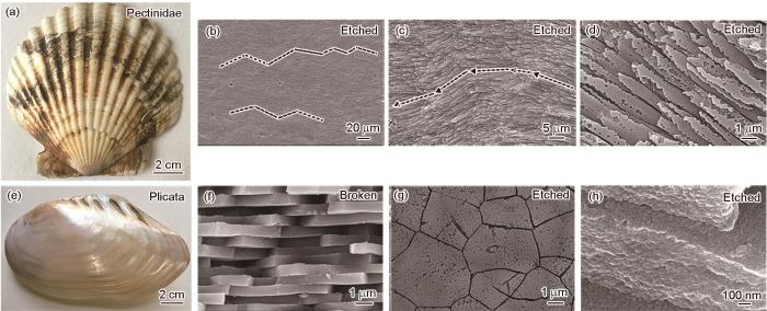
图1
扇贝的宏观形貌、腐蚀后横截面的微观结构以及褶纹冠蚌的宏观形貌、横截面自然断口形貌、贝壳平面内的腐蚀形貌、矿物板片腐蚀后的微观形貌
Fig.1
Overall views and microstructures of the Pectinidae (a~d) and Plicata (e~h) shells
1.2 观察样品的自然断口和腐蚀后的形貌
先用水冷低速金刚石切割机制备贝壳的块状材料,然后掰断以产生自然断口。依次用600#、1200#和2000#的砂纸将用于腐蚀的样品仔细研磨,用0.5#金刚石研磨膏机械抛光后将其在0.1 mol/L的乙二胺四乙酸(EDTA)溶液中腐蚀1 min。用无水乙醇充分清洗自然断口和腐蚀样品,在Ultra Plus冷场发射电子显微镜(SEM)下观察其形貌。
1.3 生物活性样品的制备
用金刚石线切割机将原贝壳切成尺寸为0.8 cm×2 cm、厚度为0.7 mm的块状样品,依次用粗细金相砂纸将表面细磨至光滑。将样品进行超声清洗后,放入烘箱中干燥1 h。
1.4 贝壳体外生物活性的测试
用磷酸氢二钠(Na2HPO4·12H2O)和磷酸(H3PO4)分别配制0.2 mol/L Na2HPO4和5 g/L H3PO4溶液。将Na2HPO4溶液滴定到H3PO4溶液中得到pH值为7.4的磷酸缓冲溶液(PBS)。用磷酸缓冲溶液浸泡法对两种贝壳的片层进行预处理,然后将其置于37℃的人体模拟体液(142.0 mmol/L Na+,5.0 mmol/L K+,1.5 mmol/L Mg2+,2.5 mmol/L Ca2+,148.8 mmol/L Cl-,4.2 mmol/L HCO
第一阶段(PBS浸泡):将每种贝壳的6个生物活性样品分成三组,第一、二组有1个扇贝样品和1个褶纹冠蚌贝壳样品,第三组有4个扇贝样品和4个褶纹冠蚌贝壳样品。将三组样品都放入12个盛有PBS溶液的塑料瓶中浸泡,第一组浸泡1 d,第二组浸泡3 d,第三组浸泡5 d。将标记的塑料瓶放入37℃的恒温振荡器中。达到浸泡时间后,将样品依次用蒸馏水、无水乙醇清洗后置于烘箱干燥皿中干燥,并标记贝壳的种类和浸泡时间。
第二阶段(SBF浸泡):从第一阶段浸泡时间为5 d的第三组样品中取3个扇贝样品和3个褶纹冠蚌贝壳样品,再将这6个样品分成三组,每组各有1个扇贝样品和1个褶纹冠蚌贝壳样品。将三组样品放入6个盛有SBF溶液的塑料瓶中浸泡,浸泡时间分别为5、10和15 d。在小塑料瓶上标记样品的种类和浸泡天数后放入37℃的恒温震荡器。达到浸泡时间后,重复阶段一中对样品的后续处理。
1.5 样品的表征
用D/MAX-RB型X射线衍射仪(XRD)分析样品的物相:Cu-K辐射(λ=0.15406 nm),加速电压40 kV,电流50 mA,扫描速率10 (°)/min,扫描范围为θ=20°~80°。用型号为4300 DV(ICP-AES)的电感耦合等离子体原子发射光谱检测浸泡后PBS溶液中离子浓度的变化,用PHS-3E电极检测PBS溶液pH值的变化。
2 实验结果
2.1 扇贝和褶纹冠蚌的微观结构
2.2 贝壳浸泡在PBS中磷灰石在其表面的生长
图2给出了两种贝壳的原始样品及其在PBS浸泡不同时间后的XRD谱。由图2可见,扇贝中的无机相是方解石型碳酸钙,而褶纹冠蚌的无机相是文石型碳酸钙。图2a给出了扇贝样品浸泡在磷酸缓冲(PBS)液中前后的XRD谱。可以看出,在PBS液中浸泡1 d后磷灰石的特征峰强度较弱,浸泡3 d后磷灰石特征峰逐渐增强,表明更多的扇贝表层转化成磷灰石;浸泡5 d后出现了新的磷灰石特征峰(2θ=32.9°),说明表层磷灰石的含量逐渐提高。图2b给出了褶纹冠蚌样品在PBS溶液中浸泡前后的XRD谱。可以看出,浸泡1 d后谱中出现了磷灰石的特征峰,说明样品表层有磷灰石生成,但是峰强远小于褶纹冠蚌原始相对应峰的峰强;浸泡3 d和5 d后磷灰石的特征峰的强度提高,但是没有出现新的特征峰,表明磷灰石的转化效率较低,也表明生成的磷灰石结晶度较低[19]。
图2
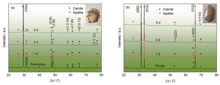
图2
扇贝和褶纹冠蚌贝壳样品浸泡PBS前后的XRD谱
Fig.2
XRD patterns of the Pectinidae (a) and Plicata (b) shell samples before and after soaking in PBS
图3
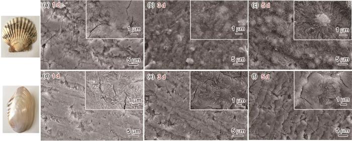
图3
扇贝和褶纹冠蚌贝壳样品浸泡在PBS后的SEM照片
Fig. 3
SEM images showing the surface morphologies of Pectinidae (a~c) and Plicata (d~f) shell samplessoaked in PBS for 1, 3 and 5 days, respectively
与褶纹冠蚌相比,在相同的条件下,在具有交叉叠片结构的扇贝上生成的磷灰石数量较多且比较致密。
图4给出了两种贝壳样品的X射线能谱(EDS),分析了样品微区的元素含量和表面生成物的Ca/P比。可以看出,在PBS中浸泡后贝壳表面发生了溶解反应而游离出钙离子。溶液中的离子浓度达到过饱和时,钙离子与溶液中的磷酸根离子反应生成片状的磷酸钙盐并沉积到贝壳表面。从图4给出的EDS数据表明,样品在PBS中浸泡1 d后表层的Ca/P比较高。其原因是,生成的磷灰石不多,数据源于磷灰石与方解石或文石的混合物。浸泡3 d后两种贝壳的Ca/P比都明显降低,表明在表层上已覆盖了大面积的磷灰石。浸泡1 d和3 d后扇贝样品表面沉积物的Ca/P较小,表明扇贝样品表面的沉积速率高于褶纹冠蚌样品。浸泡5 d后两种贝壳的Ca/P比趋于稳定,约为1.4。标准羟基磷灰石的Ca/P比为1.67,表明在PBS中浸泡后生成的是缺钙型羟基磷灰石。
图4
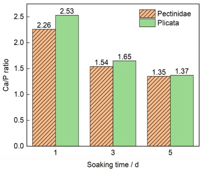
图4
扇贝和褶纹冠蚌贝壳样品在PBS中浸泡后表面Ca/P的变化
Fig.4
Changes in Ca/P ratio of Pectinideaand Plicata shell samples after soaking in PBS
碳酸钙分解后产生的钙离子,使溶液的钙离子浓度提高。由图5可见,浸泡扇贝的PBS中钙离子的浓度高于浸泡的褶纹冠蚌的PBS,表明在相同的时间内扇贝表层发生的分解反应速度更高。而同在这个阶段,两种贝壳浸泡后PBS的pH值都增大,因为溶液中发生了水解反应
图5
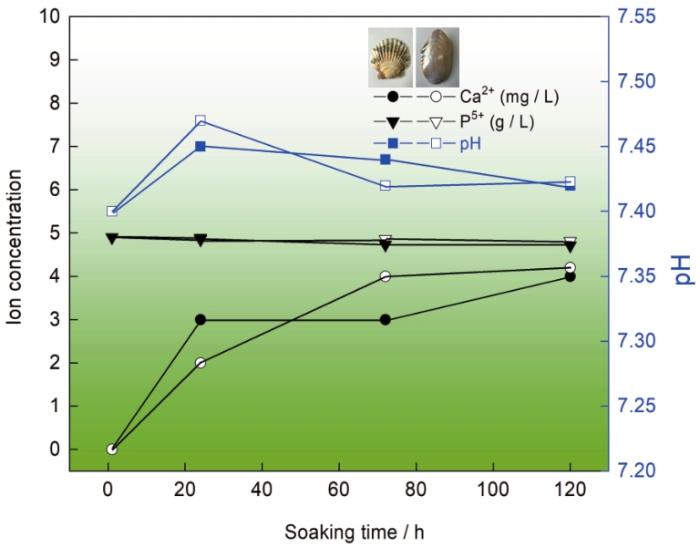
图5
扇贝和褶纹冠蚌贝壳样品在PBS中浸泡不同时间后溶液中Ca2+与P5+浓度和pH值的变化
Fig.5
Changes of Ca2+ and P5+ concentrations and pH value of Pectinidea and Plicata shell samples after soaking in PBS for different time
CO
其中0≤x<2。生成的磷灰石沉积在贝壳的表层,分解反应产生钙离子的速度略高于生成磷灰石消耗钙离子的速度,使溶液中的钙离子浓度略有提高。而在此阶段,浸泡两组样品溶液中的pH值均下降,因为磷酸根发生了分步水解
和
这是此阶段主要的水解反应,溶液中氢离子的浓度提高而溶液的pH值下降。浸泡5 d后的废液中各离子的浓度表明,发生的主要反应为
根据以上分析,钙离子在磷灰石结晶过程中发挥了重要作用,而PBS中的钙离子浓度的高低取决于生物文石的溶解。很明显,两种不同的微观结构表现出不同的溶解度:在相同的时间内扇贝表层发生的分解反应速度更高,因为贝壳的结构和无机物的组成都不同。
2.3 贝壳浸泡在SBF溶液中磷灰石在其表面的生长
图6
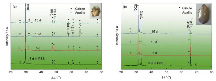
图6
扇贝和褶纹冠蚌贝壳样品在PBS预处理后在SBF溶液中浸泡5、10和15 d后的XRD谱
Fig.6
XRD patterns ofPectinidea (a) and Plicata (b) shell samples firstly treated in PBS for 5 d and then soaked in SBF for different times
图7
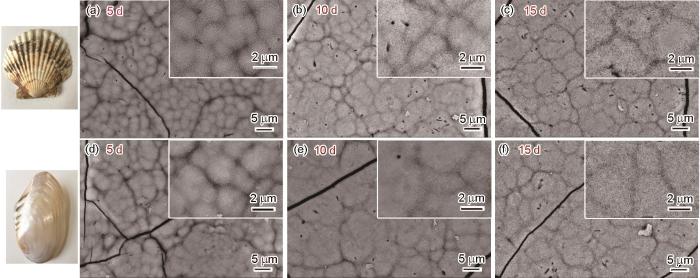
图7
用PBS预处理后扇贝和褶纹冠蚌贝壳样品在SBF溶液中浸泡5、10和15 d的SEM照片
Fig.7
SEM images showing the surface morphologies of Pectinidea (a~c) and Plicata (d~f) shell samples soaked in SBF for 5, 10 and 15 d, respectively
对比处理时间相同的扇贝和褶纹冠蚌可见,磷灰石晶体在扇贝表面生长的速度更高,磷灰石层的厚度也增加得更多,最终填满片状磷灰石间的空隙。这表明,扇贝的体外生物活性比褶纹冠蚌更好。
贝壳在PBS溶液中浸泡后,在其上生成的磷灰石为缺钙型磷灰石。晶格中缺少钙离子,使PO
图8
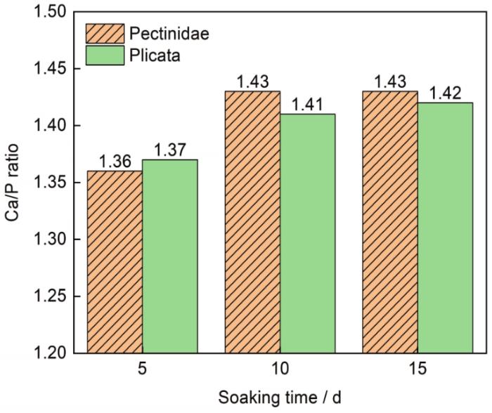
图8
浸泡在SBF中扇贝和褶纹冠蚌贝壳样品表面的Ca/P比值
Fig.8
Changes in Ca/P ratio of Pectinidae and Plicata shell samples after soaking in SBF
这表明,在浸泡的短时间内在扇贝表面生成磷灰石晶体的速度更高,磷灰石层的厚度增加得更多。但是,长时间浸泡后在两种结构表面生成的磷灰石层没有太大的差异。因此,交叉叠片结构在短期内具有较好的生物活性,可替换生物材料用于快速修复硬组织的仿生结构设计。
3 结论
(1) 扇贝具有方解石晶型交叉叠片结构,褶纹冠蚌贝壳具有文石晶型珍珠质结构。扇贝和珍珠蚌贝壳在PBS中浸泡的初始阶段,将方解石或文石(碳酸钙)转化为磷灰石的能力不同。在具有交错层状结构的扇贝表面沉积的磷灰石更紧密,沉积较快。
(2) 在SBF中的长期体外矿化过程中,在短期内扇贝具有较好的体外生物活性,在长期内两种结构都表现出优异的生物活性。
参考文献
The shell structure of the mollusks
[J].
Bivalve shell structure and organic matrix
[J].
Structural and mechanical characterization of thermally treated conch shells
[J].
Nanoscale structural and mechanical characterization of heat treated nacre
[J].
The structure and mechanical Properties of mollusk shell
[J].
香螺壳的结构和微观力学性能
[J].
Comparison of nacre with other ceramic composites
[J].
Natural arrangement of fiber-like aragonites and its impact on mechanical behavior of mollusk shells: A review
[J].
Hierarchical structure and mechanical properties of nacre: a review
[J].
Mechanical properties of crossed-lamellar structures in biological shells: A review
[J].
The self-fabrication of materials in nature offers an alternate and powerful solution towards the grand challenge of designing advanced structural materials, where strength and toughness are always mutually exclusive. Crossed-lamellar structures are the most common microstructures in mollusks that are composed of aragonites and a small amount of organic materials. Such a distinctive composite structure has a fracture toughness being much higher than that of pure carbonate mineral. These structures exhibiting complex hierarchical microarchitectures that span several sub-level lamellae from microscale down to nanoscale, can be grouped into two types, i.e., platelet-like and fiber-like crossed-lamellar structures based on the shapes of basic building blocks. It has been demonstrated that these structures have a great potential to strengthen themselves during deformation. The observed underlying toughening mechanisms include microcracking, channel cracking, interlocking, uncracked-ligament bridging, aragonite fiber bridging, crack deflection and zig-zag, etc., which play vital roles in enhancing the fracture resistance of shells with the crossed-lamellar structures. The exploration and utilization of these important toughening mechanisms have attracted keen interests of materials scientists since they pave the way for the development of bio-inspired advanced composite materials for load-bearing structural applications. This article is aimed to review the characteristics of hierarchical structures and the mechanical properties of two kinds of crossed-lamellar structures, and further summarize the latest advances and biomimetic applications based on the unique crossed-lamellar structures.Copyright © 2017 Elsevier Ltd. All rights reserved.
Demonstration of the capacity of nacre to induce bone formation by human osteoblasts maintained in vitro
[J].
Reconstruction of human maxillary defects with nacre powder: histological evidence for bone regeneration
[J].
Stimulation of bone marrow cells and bone formation by nacre: in vivo and in vitro studies
[J].
Tissue responses to nacreous implants in rat femur: an in situ hybridization and histochemical study
[J].The interface of bone and aragonite nacre (Margaritifera, fresh water pearl mussel) was studied by in situ hybridization and a tartrate-resistant acid phosphatase (TRAP) histochemical assay. Columnar implants were inserted into rat femora for 4, 7, 14, 28 and 56 days. In medullary region, a burst of transient bone formation was observed, which propagated from the periphery towards the nacre implant. A fused interface of bone and nacre was observed at 14 days. Later, the new medullary bone was resorbed and bone marrow was re-established while a thin layer of bone tissue remained covering the implant surface. Expressions of collagen alpha1(I), osteocalcin, osteopontin mRNAs and TRAP in the surrounding tissue were monitored. Correlated with the histology events, a strong transient induction of collagen alpha1(I) and osteocalcin mRNAs as well as TRAP expression, exhibiting a peak signal intensity on day 7 and subsequent down-regulation after day 14 was observed. Osteopontin mRNA, in contrast, was expressed continuously. The degrading nacre surface appeared in direct contact with macrophages and multinucleated giant cells at both days 14 and 28. These cells expressed osteopontin mRNA intensively and some TRAP enzyme activity occasionally.
Hydrothermal conversion of calcite crystals to hydroxyapatite
[J].
The bone induction effect of water soluble matrix from nacre powder on human marrow stroma cells
[J].
珍珠层水溶性提取物对人骨髓基质细胞成骨性分化的诱导作用
[J].
New crystallographic relationships in biogenic aragonite: the crossed-lamellar microstructures of mollusks
[J].
Microstructure-related in vitro bioactivity of a natural ceramic of Saxidomus purpuratus shell
[J].
Solutions able to reproduce in vivo surface-structure changes in bioactive glass-ceramic A-W3
[J].
The effect of acidic amino acids on hydroxyapatite crystallization
[J].
Nacre surface transformation to hydroxyapatite in a phosphate buffer solution
[J].
The soluble matrix from Mercenaria mercenaria shell
[J].
