


在腔镜手术和部分开放手术中,输尿管支架用于引流尿液。双猪尾型输尿管支架操作简便,得到了广泛的应用。但是这种支架植入7 d后约有10%~50%的患者发生尿路感染,植入超过30 d后感染发生概率接近100%[1, 2]。同时,约一半长期植入输尿管支架的患者因支架表面结石而发生管路堵塞[3]。近年来,区段式金属输尿管支架改变了双猪尾结构,植入后与人体输尿管内壁完全贴合,支架管腔尺寸的增大使术后堵塞概率降低[4]。但是,仍然不能避免支架表面细菌生物膜导致的尿路感染及感染性结石[5]。细菌粘附速率与尿液中细菌的种类和数量有关,也与输尿管支架材料的表面性能相关。研究发现,轻微的亲水性和强烈的表面负电性(约-60 mV)是抑制细菌生物膜形成和结石沉积的最佳表面状态[6]。使用抗生素减轻支架表面生物膜的形成,是临床中普遍使用的手段[7]。常规的抗生素疗法不仅要高剂量药物,而且长期使用会产生的耐药性[8]。
为了解决输尿管支架表面粘附细菌的问题,研究人员提出表面改性的方案。Riedl等[9]发现,在支架表面涂覆具有强烈电负性的肝素,可有效抑制微生物粘附和尿液结晶沉积。在支架表面载有酮咯酸等抗炎性药物,也是较为常用的方法[10]。动物实验结果表明,这种支架植入动物体内后感染发生率降低,输尿管和膀胱周围组织中的高浓度酮咯酸对组织和各个器官没有副作用。临床试验发现,276名患者随机接受双猪尾管支架与包被酮咯酸的支架,后者术后需要相关抗感染药物治疗的概率明显降低[11]。抗菌多肽在细菌的细胞膜上形成离子性通道,导致膜结构破裂并引起细菌胞内物质渗出,最终使细菌死亡[12]。因此,Mishra等[13]将多肽直接固定在硅树脂输尿管表面。体外研究发现,多肽对革兰氏阴性菌和革兰氏阳性菌的抗菌能力长达4 d。聚乙烯亚胺梳状高分子结构在聚氨酯表面也有持久的抗菌作用。对大鼠的体内研究发现,干扰细胞膜的信号传导可有效抑制变形奇异杆菌粘附和结石的形成[14]。此外,两性离子聚合物改性涂层构建水合层,可延迟甚至阻止微生物在材料表面粘附[15]。Wang等[16]在钛合金表面接枝两性离子聚多巴胺甲基丙烯酰胺磷酸胆碱聚合物,在满足良好生物相容性的前提下能有效地抑制细菌粘附。
在材料中引入铜元素,可使其具有抗菌性能[17]。Ren等[18]研究发现,钛铜合金在体内具有良好的生物相容性,还具有优异的抗感染功能;含铜钴铬合金支架植入小型猪的冠脉,可促进内皮化进程有效抑制术后再狭窄[19]。316L型含铜不锈钢在尿液中释放铜离子,表现出一定的抗感染和抗结石能力[20]。在新西兰白兔输尿管内植入含铜不锈钢支架80 d后,支架表面结石沉积量比316L不锈钢对照支架有所降低[21]。多巴胺在任何形状的材料表面均有极强的粘附能力,在碱性环境下多巴胺分子发生自聚合反应在材料表面形成牢固的聚多巴胺膜层。这种膜层表面有大量的苯环共轭体系,可作为金属离子的接枝反应平台[22]。本文采用聚多巴胺接枝化学镀铜方法在316L不锈钢表面制备载铜聚多巴胺涂层,研究其抗菌性能和对表面结石形成能力的抑制。
1 实验方法
1.1 实验用材料
实验用材料有:医用级316L奥氏体不锈钢,片状样品的直径为10 mm、厚度为1 mm;多巴胺盐酸盐、三羟甲基氨基甲烷(Tris)、二甲基氨硼烷(DMAB)、氯化铜、氯化钠、硫酸镁、磷酸二氢钾、磷酸二氢钠、硫酸钠、氯化钾、氯化钙和柠檬酸三钠,都为分析纯;琼脂、蛋白胨、牛肉膏;金黄色葡萄球菌冻干粉、革兰氏阴性大肠杆菌冻干粉;人输尿管上皮细胞;高糖培养基(DMEM),胎牛血清(FBS),胰蛋白酶,Hyclone;青链霉素,Logan;MTS,[3-(4,5-二甲基噻唑-2-基)-5-(3-羧甲酯基)-2-(4-磺苯基)-2H四唑(金翁),内盐],Abcam。
1.2 载铜聚多巴胺涂层样品的制备
将316L不锈钢用600#、1000#、2000#砂纸打磨后依次用去离子水、无水乙醇超声清洗2 min,干燥后备用。将316L不锈钢样品浸泡在多巴胺浓度为2 g/L的Tris-HCl缓冲溶液中(pH=8.5),避光反应24 h。然后用去离子水、无水乙醇依次超声清洗2 min,干燥后得到载有聚多巴胺涂层的316L不锈钢样品。再用化学接枝手段与CuCl2浓度为2 mol/L、DMAB浓度为0.1 mol/L的化学镀铜液在25℃条件下反应12 h,以吸附溶液中的铜单质形成载铜聚多巴胺涂层。最后将样品用去离子水、无水乙醇清洗,干燥后保存。
1.3 载铜聚多巴胺涂层性能的表征
用扫描电镜(SEM,KYKY-EM8100F,USA)观察载铜聚多巴胺涂层形貌,观察前需要进行喷金处理。用原子力显微镜(AFM,BRUKER Dimension Icon,USA),采用非接触式测量方法检测载铜聚多巴胺涂层、聚多巴胺涂层及316L不锈钢样品表面。使用Gwyddion软件处理AFM图像。
使用椭圆偏振光谱仪(J.A. Wollam, M-2000,USA)测试材料表面涂层厚度。为了提高检测精度,将316L不锈钢替换为硅片,后续涂层制备方法相同。
用X射线光电子能谱仪(XPS,Thermo VG,USA)测试载铜聚多巴胺涂层。使用ESCALAB-250俄歇表面测量系统,并使用XPSPEAK41软件对铜元素的谱图进行分峰拟合处理。
| Component | NaCl | NaH2PO4 | Na3C6H5O7 | MgSO4 | NaSO4 | KCl | Na2C2O4 | CaCl2 |
|---|---|---|---|---|---|---|---|---|
| Quantity/g | 6.17 | 4.59 | 0.944 | 0.463 | 2.408 | 4.75 | 0.043 | 0.638 |
1.4 抗菌性能的测试
采用直接接触实验法进行体外抗菌性能测试。将载铜聚多巴胺样品和316L不锈钢样品置于24孔板中,在每个样品表面滴加50 μL细菌悬液并将其均匀铺展。所用细菌悬液介质为人工尿液,细菌为金黄色葡萄球菌、大肠杆菌,细菌悬液浓度分别为5.2×10⁵、6.7×10⁵ cfu/mL。在37℃培养箱中培养24 h后将每个样品置于5 mL PBS溶液中,涡旋洗脱1 min。取100 μL稀释后的悬液均匀地涂抹在固体培养基上,在37℃培养箱中培养24 h后进行菌落计数。样品的杀菌率为
式中A1为阴性组活菌数,A2为实验组活菌数。
为了直观评价涂层的牢固度,将载铜聚多巴胺样品在生理盐水中震荡浸泡30 d。取出用去离子水清洗干燥后,用上述实验方法验证抗菌性能。
1.5 表面结石沉积实验
将样品垂直置于 15 mL 离心管中。向每个离心管中加入10 mL初始浓度为4.9×10⁷ cfu/mL的金黄色葡萄球菌悬液,细菌悬液介质为人工尿液与人体尿液1:1配制[20],在37℃恒温箱中进行无机盐沉积实验。为了模拟人体环境,每天更换人工尿液。沉积 30 d后将样品取出,用蒸馏水轻轻清洗以去除未沉积到样品表面的无机盐。将无机盐沉积后的样品置于5%(体积分数)浓度 HCl 溶液中,超声溶解5 min后用ICP-MS检测溶液中钙离子和镁离子的含量。
1.6 细胞毒性的测试
使用 MTS试剂检测细胞的毒性。依照GB/T 16686.6-2003要求,将样品在培养基中浸泡72 h。将培养瓶中的输尿管上皮细胞用胰蛋白酶消化后配制成浓度为5×10⁴ cell/mL的细胞悬液,并将100 μL加入到96孔板中。在5% CO2培养箱中培养至细胞贴壁后更换100 μL材料浸提液,同时设置阴性对照组(完全培养基)和阳性对照组(含有10% DMSO的完全培养基),分别培养24、48和 72 h。到达既定时间点取出96孔板,加入20 μL MTS,继续培养4 h,然后在酶标仪490 nm波长下测量吸光度值,细胞相对增殖率为
1.7 统计学分析
使用SPSS 20.0统计学软件进行统计学分析。每种材料测试多于3个样品,其结果用平均值±标准差(X±SD)表示。用Student’s t-test方法进行比较,*p<0.05为有显著统计学差异,△p≥0.05无显著统计学差异。
2 实验结果
2.1 载铜聚多巴胺涂层的性能
2.1.1 载铜聚多巴胺涂层的表面形貌
图1
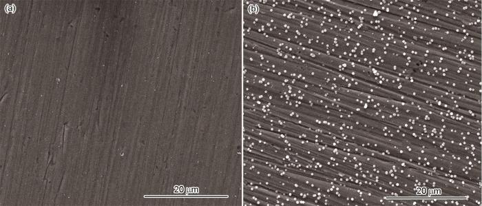
图1
316L不锈钢和载铜聚多巴胺涂层样品表面的形貌
Fig.1
SEM images of samples (a) 316L stainless steel, (b) copper loaded polydopamine coating
图2

图2
载铜聚多巴胺涂层的面扫图
Fig.2
Surface scanning images of copper loaded polydopamine coating (a) Fe element, (b) Cu element, (c) O element and (d) C element
图3
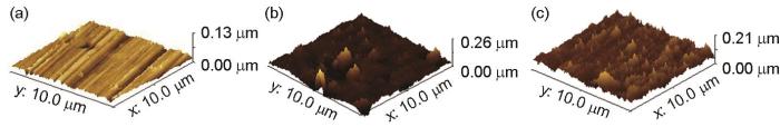
图3
316L不锈钢、聚多巴胺涂层和载铜聚多巴胺涂层样品的AFM形貌
Fig.3
AFM morphology of samples (a) 316L stainless steel, (b) polydopamine coating, (c) copper loaded polydopamine coating
2.1.2 载铜聚多巴胺涂层的膜厚结果
图4
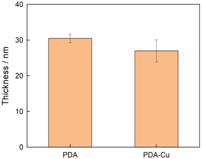
图4
用椭圆偏振光谱仪检测样品的膜厚
Fig.4
Thickness of coating measured by spectroscopic ellipsometer
2.1.3 载铜聚多巴胺涂层表面的成分
为了研究载铜聚多巴胺涂层表面的成分,分析了样品表面的XPS全谱和精细谱。图5a给出了溅射40 s的聚多巴胺涂层样品和载铜聚多巴胺涂层样品表面的XPS全谱。以C1s=284.9 eV为基准校正结合能,聚多巴胺涂层在结合能399.8 eV处出现了N1s特征峰,表明基体表面有N元素。对N1s进行分峰处理,可将其曲线拟合成两个峰,其一是结合能为399.5 eV的-N-H(氨基)峰,其二是结合能为398.5 eV的=N-(亚氨基)峰,如图5b所示。-N-H源于多巴胺结构中的氨基基团,而=N-是在多巴胺氧化自聚合过程中吲哚结构异变形成的,表明聚多巴胺已经粘附在316L不锈钢表面。为了验证这一论证,进一步分析了C1s高分辨XPS图谱。C1s分峰处理后,主要包含C-C/C-H(284.8 eV)、C-N(285.4 eV)和C-N/C-O(286 eV)3个峰,上述特征峰的出现,也表明聚多巴胺沉积在316L不锈钢表面。此外,根据聚多巴胺涂层表面的XPS定量计算出N/C比值为0.121,与多巴胺结构的理论值0.125较为接近。这进一步证明,多巴胺已经自聚合在316L不锈钢材料表面[25]。载铜聚合多巴胺涂层在结合能952.4 eV处出现了Cu2p的特征峰,表明基体表面装载了铜元素。为进一步分析涂层表面所载铜元素的成分,对溅射40 s的铜的特征峰进行分峰处理,分解出Cu(标准峰为932.4 eV)、CuO(标准峰为933.6 eV)、Cu2O(标准峰为932 eV)三个主峰,如图5b所示。根据分峰后主峰的面积比计算出溅射不同时间三种铜元素形成物质的百分含量,结果列于表2。溅射40、60、80 s时Cu、CuO与Cu2O的质量百分比呈增加趋势,表明涂层中铜的分布有一定的梯度,存在水溶液时外层的铜优先发生氧化反应并以离子的形式释放。
图5
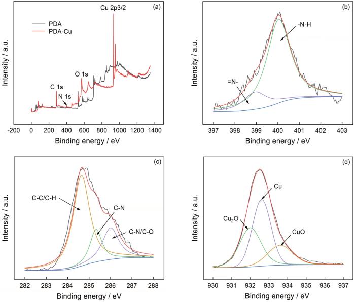
图5
载铜聚多巴胺涂层的XPS分析结果
Fig.5
XPS analysis of copper loaded polydopamine coating (a) full spectrum, (b) N element, (c) C element and (d) Cu element
表2 溅射不同时间的载铜聚多巴胺涂层中Cu、CuO和Cu2O的含量
Table 2
| Sputtering time/s | Cu | CuO | Cu2O |
|---|---|---|---|
| 40 | 2.9 | 0.8 | 1.5 |
| 60 | 4.5 | 2.6 | 3.4 |
| 80 | 7.5 | 3.9 | 5.4 |
2.2 铜离子的溶出
图6
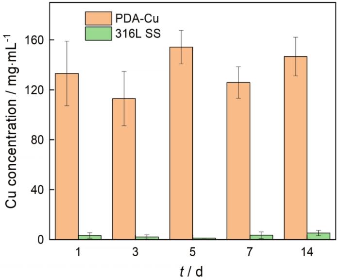
图6
不同样品在人工尿液中浸泡后每天释放的铜离子含量
Fig.6
Release amounts of Cu ions of different samples in artificial urine for every day
2.3 抗菌性能
图7
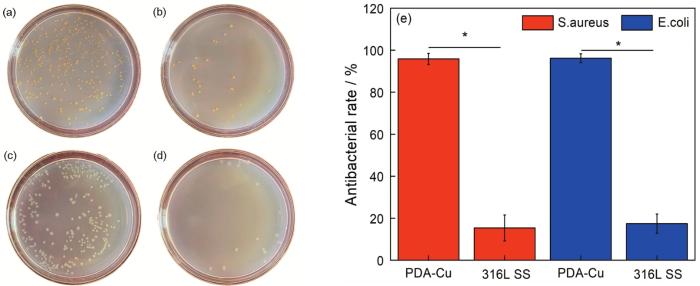
图7
不同材料与细菌培养24 h后的活细菌数量和抗菌率
Fig.7
Live bacteria number and antibacterial rate of different samples after incubation for 24 h (a) S.aureus, 316L SS; (b) S.aureus, copper loaded polydopamine coating; (c) E.coli, 316L SS; (d) E.coli, copper loaded polydopamine coating; (e) antibacterial rate
图8
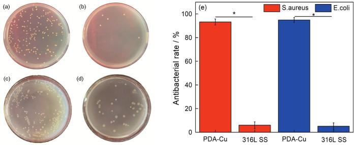
图8
材料与细菌浸泡培养30 d后活细菌的数量和抗菌率
Fig.8
Live bacteria number and antibacterial rate of different samples after immersion for 30 d (a) S.aureus, 316L SS; (b) S.aureus, copper loaded polydopamine coating; (c) E.coli, 316L SS; (d) E.coli, copper loaded polydopamine coating; (e) antibacterial rate
2.4 抗结石性能
结石的主要成分是钙盐和镁盐。图9给出了在316L不锈钢与载铜聚多巴胺涂层样品表面沉积的结石中钙、镁离子的含量。316L不锈钢载铜聚多巴胺涂层样品和316L不锈钢样品表面沉积的钙离子含量分别为48.7 mg/L和66.9 mg/L;镁离子含量分别为235.3 mg/L和342.6 mg/L,均具有显著的统计学差异。
图9

图9
不同材料在尿液中沉积30 d后表面结石中钙、镁离子的含量
Fig.9
Contents of calcium (a) and magnesium (b) ions on the surface of different materials after immersion in urine for 30 d
2.5 涂层的细胞毒性
为了验证载铜聚多巴胺涂层在泌尿系统中生物安全性,将材料浸提液与输尿管上皮细胞(UECs)共培养24、48及72 h,MTS测试结果如图10所示。可以看出,随着培养时间的增加输尿管的上皮细胞与316L不锈钢、聚多巴胺涂层及载铜聚多巴胺涂层培养后的相对增殖率均高于90%,毒性评价为1级,符合GB/T 16886.5-2017《医疗器械生物学评价——第五部分,体外细胞毒性试验》对植入材料毒性的要求。
图10

图10
UECs与不同材料浸提液培养不同时间后的相对增殖率
Fig.10
Relative proliferation rate of UECs cultured with different material extracts for different times
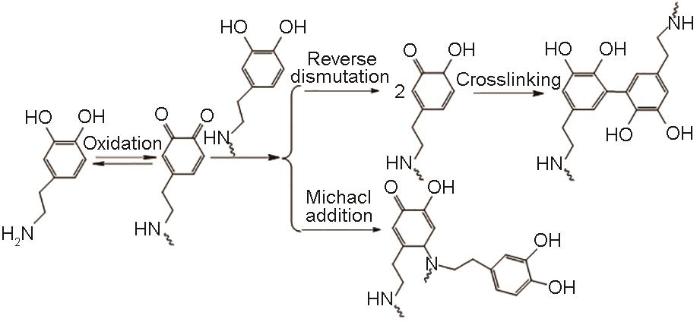
3 讨论
多巴胺在不同材料及不同形状样品上具有强力粘附的特性,本文用多巴胺将316L不锈钢与铜结合。图11给出了多巴胺的自聚合反应式[26]。溶液中的氧和碱性条件使多巴胺的邻苯二酚氧化成结构不稳定的多巴胺醌,多巴胺醌发生分子内环化后形成多巴胺中间体,再进行氧化重排、交联形成聚多巴胺[27]。316L不锈钢经多巴胺处理后,多巴胺发生聚合反应在其表面形成稳定的聚多巴胺涂层。在化学镀铜液中铜离子被还原剂二甲基硼氨烷还原成铜单质,吸附在聚多巴胺涂层上。扫描电镜和原子力显微镜结果表明,聚多巴胺在316L不锈钢表面的粘附使316L不锈钢表面的微观结构发生变化。同时,聚多巴胺均匀地吸附了铜,图5给出的XPS结果中铜以Cu、CuO和Cu2O的形式存在,在人体尿液环境中发生氧化还原反应实现了铜离子的释放。
图11
Cu+是铜发挥杀菌作用的主要离子,通过破坏细菌中的Fe-S蛋白与硫元素结合形成了Cu+-S,从而杀灭细菌[28]。但是,溶液中的Cu+稳定性差,水合焓为-593 kJ/mol;而Cu2+则较稳定,其水合焓为-2099 kJ/mol。因此,在水溶液中Cu+浓度较低,易发生歧化反应形成Cu2+和Cu[29, 30]。这表明,提高Cu+浓度是提高涂层抗菌性能的重要途径。不同于单纯利用多巴胺螯合Cu2+的载铜制备方法,本文用多巴胺化学接枝镀铜方法在材料表面制备富有Cu及其氧化物的混合载铜涂层,通过铜元素不同价态间的反应实现抗菌功能。载铜聚多巴胺涂层与空气中的氧气和水接触后发生氧化反应生成Cu2O和CuO混合的铜的氧化物,反应式为
Cu2O和CuO不断与人体尿液中的H+反应生成Cu2+,反应式为
尿液中的Cu+、Cu2+和Cu在不断反应中达到平衡,反应式即
载铜聚多巴胺涂层通过铜的系列反应实现了表面改性的功能,使其具有生物功能性。
4 结论
用多巴胺化学接枝镀铜方法可在316L不锈钢表面制备聚多巴胺涂层。通过化学接枝手段,在化学镀铜液中铜离子被还原剂二甲基硼氨烷还原成铜单质,吸附在聚多巴胺涂层形成载铜聚多巴胺涂层。载铜聚多巴胺涂层中的铜以Cu、CuO和Cu2O的形式存在,在人工尿液中不断释放以离子,表现出良好的杀灭大肠杆菌和金黄色葡萄球菌的性能。通过抑制尿液中的细菌进一步降低样品表面晶体沉积,表现出良好的抗结石能力。载铜聚多巴胺涂层没有细胞毒性,可用于输尿管支架。
参考文献
Observation of double J stent bacterial colonisation and its correlation with bacteriuria frequency
[J].
In vitro effects of a novel coating agent on bacterial biofilm development on ureteral stents
[J].
Ureteral stent encrustation: epidemiology, pathophysiology, management and current technology
[J].
Allium stent for the treatment of a malignant ureteral stenosis: a paradigmatic case
[J].
Long-term outcomes of two types of metal stent for chronic benign ureteral strictures
[J].
We aimed to compare the results of long-term use of two types of metal stent for chronic benign ureteral strictures.Our study included 46 ureter units (UUs) that underwent metal stent placement from 2010 to 2017. We included benign ureteral strictures causes by variety reasons that could not be solved by other treatment and malignant obstructions were excluded. Covered mesh stent (Uventa™) and a thermo-expandable stent (Memokath 051™) were used. Primary success was defined as maintaining patency without procedures and overall success was defined as maintaining patency with additional procedures.We placed covered mesh stents in 25 UUs and thermo-expandable stents in 21 UUs. The mean follow-up duration of each stent was 41.4 ± 23.1 and 34.4 ± 16.5 months (p = 0.250). In the first year of stent insertion, primary success was achieved in 54.9 and 70.4% (p = 0.204). Overall success was achieved in 78.7 and 75.4% in same duration, respectively (p = 0.586). Longer stent placement had positive predictive value on both success rates (HR = 0.185, p = 0.047 and HR = 0.111, p = 0.018). Prior radiation therapy and non-pelvic ureter stricture both adversely affected the overall success rate (HR = 5.412, p = 0.048 and HR = 4.203, p = 0.030). Previous PCN status had negative predictive value for both success rates (HR = 4.014, p = 0.003 and HR = 3.064, p = 0.035).The treatment outcomes of two types of metal stent were comparable, especially in the first year of stent insertion.
Prevention of encrustation on ureteral stents: which surface parameters provide guidance for the development of novel stent materials?
[J].
Strategies for combating bacterial biofilms: a focus on anti-biofilm agents and their mechanisms of action
[J].Biofilm refers to the complex, sessile communities of microbes found either attached to a surface or buried firmly in an extracellular matrix as aggregates. The biofilm matrix surrounding bacteria makes them tolerant to harsh conditions and resistant to antibacterial treatments. Moreover, the biofilms are responsible for causing a broad range of chronic diseases and due to the emergence of antibiotic resistance in bacteria it has really become difficult to treat them with efficacy. Furthermore, the antibiotics available till date are ineffective for treating these biofilm related infections due to their higher values of minimum inhibitory concentration (MIC) and minimum bactericidal concentration (MBC), which may result in in-vivo toxicity. Hence, it is critically important to design or screen anti-biofilm molecules that can effectively minimize and eradicate biofilm related infections. In the present article, we have highlighted the mechanism of biofilm formation with reference to different models and various methods used for biofilm detection. A major focus has been put on various anti-biofilm molecules discovered or tested till date which may include herbal active compounds, chelating agents, peptide antibiotics, lantibiotics and synthetic chemical compounds along with their structures, mechanism of action and their respective MICs, MBCs, minimum biofilm inhibitory concentrations (MBICs) as well as the half maximal inhibitory concentration (IC) values available in the literature so far. Different mode of action of anti biofilm molecules addressed here are inhibition via interference in the quorum sensing pathways, adhesion mechanism, disruption of extracellular DNA, protein, lipopolysaccharides, exopolysaccharides and secondary messengers involved in various signaling pathways. From this study, we conclude that the molecules considered here might be used to treat biofilm-associated infections after significant structural modifications, thereby investigating its effective delivery in the host. It should also be ensured that minimum effective concentration of these molecules must be capable of eradicating biofilm infections with maximum potency without posing any adverse side effects on the host.
Engineering solutions to ureteral stents: material, coating and design
[J].
Heparin coating reduces encrustation of ureteral stents: a preliminary report
[J].
An in vivo porcine evaluation of the safety, bioavailability, and tissue penetration of a ketorolac drug-eluting ureteral stent designed to improve comfort
[J].Ureteral stents often cause significant patient morbidity that can be difficult to treat. Drug-eluting stent technology allows the local delivery of a drug. Our previous work demonstrated that ketorolac instilled intravesically at the time of ureteral stent insertion significantly decreased flank pain compared with controls. We sought to determine the safety of a novel ketorolac-eluting ureteral stent.A total of 92 Yorkshire pigs were randomized to 1 of 5 groups. The oral control group consisted of 12 animals with transurethrally inserted control ureteral stents and 5 days of oral ketorolac. Twenty animals in each of the remaining groups received a control stent, or 15%, 13%, or 7% ketorolac-loaded stents. Ketorolac levels were measured in plasma, urine, and tissue sampled from ureters, bladder, kidneys, and liver using high performance liquid chromatography. Necropsies were performed to evaluate tissue pathology.The majority of ketorolac was released within the first 30 days. The highest levels of ketorolac in plasma, kidney, and liver occurred in the oral control group. The highest levels of ketorolac found in ureteral and bladder tissues occurred in the ketorolac-stent groups in a dose-dependent fashion. No adverse events were noted in any of the ketorolac-stent groups. Gastric ulcerations were identified only in the oral control group. No abnormalities were identified in any other internal organs in any group.The use of ketorolac-eluting ureteral stents has proven to be safe in a porcine model. The ketorolac-stent group had less than 12% of the ketorolac concentration in plasma, kidney, and liver tissues compared with the oral ketorolac group. Ureteral tissues displayed the highest levels of ketorolac. Clinical studies are needed to determine if ketorolac-elution reduces stent symptoms.
A novel drug eluting ureteral stent: a prospective, randomized, multicenter clinical trial to evaluate the safety and effectiveness of a ketorolac loaded ureteral stent
[J].We evaluated the short-term safety and efficacy of a ketorolac loaded ureteral stent compared to a standard stent (control).In this prospective, multicenter, double-blind study patients were randomized 1:1 to ketorolac loaded or control stents after ureteroscopy. The primary end point was an intervention for pain defined as unscheduled physician contact, change in pain medication or early stent removal. Secondary end points included medication use and pain visual analog score. A total of 20 patients underwent serum safety testing for ketorolac levels.None of the safety cohort had detectable serum ketorolac levels. Among the 276 patients there was no difference in primary (9.0% ketorolac loaded vs 7.0% control, p = 0.66) or secondary (22.6% ketorolac loaded vs 25.2% control, p = 0.67) intervention rates. Mean pain pill count at day 3 was lower in the ketorolac loaded stent group than in the control group (p <0.05). A higher number (p = 0.057) of patients with ketorolac loaded (32%) stents used no or limited pain medications compared to controls (22%). A higher number of male patients with ketorolac loaded stents used no pain medication on days 3 and 4 compared to female patients with ketorolac loaded stents, and male and female control patients (p <0.05).The overall safety of the ketorolac loaded stent was confirmed. Although there was no significant difference in primary or secondary intervention rates, a trend toward a treatment benefit was noted for patients receiving drug loaded stents. Specifically young male patients appeared to require less pain medication when the ketorolac loaded stent was used. Future studies with higher drug concentrations or alternative drug eluting stents may prove beneficial.2010 American Urological Association Education and Research, Inc. Published by Elsevier Inc. All rights reserved.
Polyalanine peptide variations may have different mechanisms of action against multidrug-resistant bacterial pathogens
[J].The number of bacterial pathogens resistant to the currently available antibiotics has dramatically increased, with antimicrobial peptides (AMPs) being among the most promising potential new drugs. In this study, the applicability and mechanisms of action of Pa-MAP 2 and Pa-MAP 1.9, two AMPs synthetically designed based on a natural AMP template, were evaluated.Pa-MAP 2 and Pa-MAP 1.9 were tested against a clinically isolated multidrug-resistant (MDR) Escherichia coli strain. Biophysical approaches were used to evaluate the preference of both peptides for specific lipid membranes, and bacterial surface changes imaged by atomic force microscopy (AFM). The efficacy of both peptides was assessed both in vitro and in vivo.Experimental results showed that both peptides have antimicrobial activity against the E. coli MDR strain. Zeta potential and surface plasmon resonance assays showed that they interact extensively with negatively charged membranes, changing from a random coil structure, when free in solution, to an α-helical structure after membrane interaction. The antibacterial efficacy was evaluated in vitro, by several techniques, and in vivo, using a wound infection model, showing a concentration-dependent antibacterial effect. Different membrane properties were evaluated to understand the mechanism underlying peptide action, showing that both promote destabilization of the bacterial surface, as imaged by AFM, and change properties such as membrane surface and dipole potential.Despite their similarity, data indicate that the mechanisms of action of the peptides are different, with Pa-MAP 1.9 being more effective than Pa-MAP 2. These results highlight their potential use as antimicrobial agents against MDR bacteria.© The Author(s) 2021. Published by Oxford University Press on behalf of the British Society for Antimicrobial Chemotherapy. All rights reserved. For permissions, please email: journals.permissions@oup.com.
Site specific immobilization of a potent antimicrobial peptide onto silicone catheters: evaluation against urinary tract infection pathogens
[J].
Designing of dynamic polyethyleneimine (PEI) brushes on polyurethane (PU) ureteral stents to prevent infections
[J].Permanent antibacterial coatings have been developed by brush-like polyethyleneimine (PEI) on polyurethane (PU) ureteral stents since bacterial adhesion and biofilm formation with the following encrustation on stent surface limit their long term usage. In order to control or prevent bacterial infections; PEI chains with two different molecular weights (Mn: 1800 or 60,000 Da) were covalently attached on the polyurethane (PU) surface by "grafting to" approach to obtain a brush-like structure. Then, PEI brushes were alkylated with bromohexane to enhance the disruption of bacterial membranes with increasing polycationic character. X-ray Photoelectron and Infrared Spectroscopy investigations confirmed that PEI grafting and alkylation steps were performed successfully. Surface roughness in dry state increased dramatically from 65.8 nm to 277.7 nm and 145.2 nm for short chain PEI and long chain PEI grafted samples, respectively. Both low and high molecular weight PEI grafts exhibited a brush-like structure and potent antibacterial activity by lowering the adherence of Klebsiella pneumonia, Escherichia coli and Proteus mirabilis species up to two orders of magnitude without any cytotoxic effect on L929 and G/G cells. Thus, permanent bactericidal activity was achieved by the contact-active strategy of dynamic PEI brush-like structures on polyurethane ureteral stent.Copyright © 2015 Acta Materialia Inc. Published by Elsevier Ltd. All rights reserved.
Antifouling photograftable zwitterionic coatings on PDMS substrates
[J].The foreign body response (FBR) to implantable materials can negatively impact performance of medical devices such as the cochlear implant. Engineering surfaces that resist the FBR could lead to enhanced functionality including potentially improving outcomes for cochlear implant recipients through reduction in fibrosis. In this work, we coat poly(dimethylsiloxane) (PDMS) surfaces with two zwitterionic polymers, poly(sulfobetaine methacrylate) (pSBMA) and poly(carboxybetaine methacrylate) (pCBMA), using a simultaneous photografting/photo-cross-linking process to produce a robust grafted zwitterionic hydrogel. reduce nonspecific protein adsorption, the first step of the FBR. The coating process uses benzophenone, a photografting agent and type II photoinitiator, to covalently link the cross-linked zwitterionic thin film to the PDMS surface. As the concentration of benzophenone on the surface increases, the adhesive strength of the zwitterionic thin films to PDMS surfaces increases as determined by shear adhesion. Additionally, with increased concentration of the adsorbed benzophenone, failure of the system changes from adhesive delamination to cohesive failure within the hydrogel, demonstrating that durable adhesive bonds are formed from the photografting process. Interestingly, antifouling properties of the zwitterionic polymers are preserved with significantly lower levels of nonspecific protein adsorption on zwitterion hydrogel-coated samples compared to uncoated controls. Fibroblast adhesion is also dramatically reduced on coated substrates. These results show that cross-linked pSBMA and pCBMA hydrogels can be readily photografted to PDMS substrates and show promise in potentially changing the fibrotic response to implanted biomaterials.
Articular cartilage-inspired surface functionalization for enhanced lubrication
[J].
In vitro study of role of trace amount of Cu release from Cu-bearing stainless steel targeting for reduction of in-stent restenosis
[J].
Biocompatibility and Cu ions release kinetics of copper-bearing titanium alloys
[J].To reduce the risk of implant-associated infections, we previously designed and developed a series of medical copper (Cu)-bearing titanium alloys that release Cu ions and hence play an antibacterial role. However, both excessive and deficient Cu levels adversely affect human health; therefore, the aim of the present study was to comprehensively evaluate the short- and long-term biosafety of Cu-bearing titanium alloys (Ti6Al4V-Cu and Ti-Cu) both in vitro and in vivo. Moreover, the predominant kinetic mechanism of Cu ions release and its effect on biosafety were also investigated. The results indicate that the biocompatibility of the Cu-bearing titanium alloys meets the requirements of ISO standards and the Cu ion release kinetics display a good correlation over the entire time period in the normal zero-order model with an almost constant release rate. The release rate maintained at a parts per billion level safe for humans; consequently, we can conclude that our Cu-bearing titanium alloys have satisfactory biocompatibility.
In vitro study of stimulation effect on endothelialization by a copper bearing cobalt alloy
[J].
In vitro study on infectious ureteral encrustation resistance of Cu-bearing stainless steel
[J].Cu-bearing stainless steel has been found to have obvious inhibition performance against encrustation in vitro. This study was aiming to further investigate the inhibitory effect of a Cu-bearing stainless steel (316L-Cu SS) on the infectious encrustation based on its antimicrobial activity. The encrustation in presence of bacteria, antibacterial performance, urease production and Ca and Mg precipitation were examined by scanning electron microscopy, antibacterial assay, enzyme-linked immunosorbent assay and inductively coupled plasma-mass spectrometry, respectively. It was found that 316L-Cu SS could inhibit the formation of bacterial biofilm due to the release of Cu2+ ions and then decrease the urease amount splitting by bacteria, which produced a neutral environment with pH around 7. However, more encrustations coupled with bacterial biofilms on the surface of comparison stainless steel (316L SS) with an alkaline environment were recorded. It can thus be seen that the 316L-Cu SS highlights prominent superiority against encrustation in the presence of microorganisms.
In vivo research on Cu-bearing ureteral stent
[J].
A facile heparin/carboxymethyl chitosan coating mediated by polydopamine on implants for hemocompatibility and antibacterial properties
[J].
Construction of copper mediated in situ nitric oxide-generating coating for cardiovascular interventional devices
[D].
用于血管介入器械表面改性的铜介导原位催化产生一氧化氮涂层的构建
[D].
Fabrication and properties of silverized glass fiber by dopamine functionalization and electroless plating
[J].
Preparation and antibacterial function of an Cu-bearing chitosan coating on silicone rubber surface
[J].In order to solve the infection caused by the indwelling catheter, an anti-infective Cu-bearing chitosan coating was prepared on the silicone rubber surface. But it is difficult to prepare a coating on the surface of silicone rubber due to its biological inertness. Therefore, chemical grafting was used to activate the silicone rubber by the dopamine pretreatment, which provides abundant functional groups on the activated silicone rubber surface. The surface morphology and surface properties of the silicone rubber after surface activation pretreatment were characterized by the active functional groups. Onto which, subsequently, the Cu-bearing chitosan coating could be chemically grafted, and then the surface morphology was compared for the coatings before and after immersion test. The effectiveness of pretreatment process was assessed by the bonding force between the functionalized coating and the silicone rubber. It follows that the abundant functional groups offered by the pretreatment on the activated silicone rubber surface may be beneficial for enhancing the adhesive strength of the functionalized coating to the silicon rubber. Thereby, the Cu-bearing chitosan coating makes the silicone rubber catheter have good antibacterial function.
硅橡胶表面壳聚糖载铜凝胶涂层的制备及其抗菌功能
[J].在硅橡胶表面制备一种具有抗感染功能的涂层—壳聚糖载铜凝胶涂层。为了克服硅橡胶的生物惰性,在其表面制备涂层,先用逐步化学接枝法对其表面进行活化预处理,然后化学接枝壳聚糖载铜凝胶涂层。对比浸泡前后涂层的形貌,研究了活化预处理对功能化涂层与硅橡胶基体之间结合性能的影响。结果表明,用化学接枝法可在硅橡胶表面生成丰富的活性官能团从而提高了功能化涂层与硅橡胶的结合强度。载铜功能化涂层使硅橡胶导管具有良好的抗菌功能。
Construction of multilyered composites from poly (dopamine) modified carbon nanotubes and the interactions with cells
[D].
聚多巴胺修饰碳纳米管静电叠层复合膜的制备及细胞作用研究
[D].
Quantitative proteomic profiling of the Escherichia coli response to metallic copper surfaces
[J].Metallic copper surfaces have strong antimicrobial properties and kill bacteria, such as Escherichia coli, within minutes in a process called contact killing. These bacteria are exposed to acute copper stress under dry conditions which is different from chronic copper stress in growing liquid cultures. Currently, the physiological changes of E. coli during the acute contact killing process are largely unknown. Here, a label-free, quantitative proteomic approach was employed to identify the differential proteome profiles of E. coli cells after sub-lethal and lethal exposure to dry metallic copper. Of the 509 proteins identified, 110 proteins were differentially expressed after sub-lethal exposure, whereas 136 proteins had significant differences in their abundance levels after lethal exposure to copper compared to unexposed cells. A total of 210 proteins were identified only in copper-responsive proteomes. Copper surface stress coincided with increased abundance of proteins involved in secondary metabolite biosynthesis, transport and catabolism, including efflux proteins and multidrug resistance proteins. Proteins involved in translation, ribosomal structure and biogenesis functions were down-regulated after contact to metallic copper. The set of changes invoked by copper surface-exposure was diverse without a clear connection to copper ion stress but was different from that caused by exposure to stainless steel. Oxidative posttranslational modifications of proteins were observed in cells exposed to copper but also from stainless steel surfaces. However, proteins from copper stressed cells exhibited a higher degree of oxidative proline and threonine modifications.
The interaction of copper ions with Staphylococcus aureus, Pseudomonas aeruginosa, and Escherichia coli: an X-ray absorption near-edge structure (XANES) spectroscopy study
[J].
Magnesium-doped zinc oxide nanoparticles alter biofilm formation of Proteus mirabilis
[J].
Bacterial adherence to silver nitrate coated poly-L-lactic acid urological stents in vitro
[J].The purpose of this study was to see whether it is possible to prevent bacterial adherence to bioabsorbable self-reinforced L-lactic acid polymer (SR-PLLA) urological stents. The SR-PLLA stents were coated with silver nitrate blended epsilon-caprolactone/L-lactide copolymer. The adherence of five bacterial strains (Pseudomonas aeruginosa, Enterococcus faecalis, Proteus mirabilis and two strains of Escherichia coli) to coated and non-coated SR-PLLA wires were tested. It was found that silver nitrate coating prevented the adherence of bacteria (except E. faecalis) to SR-PLLA stents. The preventive effect correlated with the silver nitrate concentration. It was also found that silver nitrate coating reduced the amount of bacteria in ambient urine. In conclusion, silver nitrate coating may reduce stent-associated bacterial infections by preventing the adherence of bacteria. Further studies are needed to confirm its efficacy and safety in clinical practice.
Prevention of encrustation and blockage of urinary catheters by Proteus mirabilis via pH-triggered release of bacteriophage
[J].
Biofilms: survival mechanisms of clinically relevant microorganisms
[J].Though biofilms were first described by Antonie van Leeuwenhoek, the theory describing the biofilm process was not developed until 1978. We now understand that biofilms are universal, occurring in aquatic and industrial water systems as well as a large number of environments and medical devices relevant for public health. Using tools such as the scanning electron microscope and, more recently, the confocal laser scanning microscope, biofilm researchers now understand that biofilms are not unstructured, homogeneous deposits of cells and accumulated slime, but complex communities of surface-associated cells enclosed in a polymer matrix containing open water channels. Further studies have shown that the biofilm phenotype can be described in terms of the genes expressed by biofilm-associated cells. Microorganisms growing in a biofilm are highly resistant to antimicrobial agents by one or more mechanisms. Biofilm-associated microorganisms have been shown to be associated with several human diseases, such as native valve endocarditis and cystic fibrosis, and to colonize a wide variety of medical devices. Though epidemiologic evidence points to biofilms as a source of several infectious diseases, the exact mechanisms by which biofilm-associated microorganisms elicit disease are poorly understood. Detachment of cells or cell aggregates, production of endotoxin, increased resistance to the host immune system, and provision of a niche for the generation of resistant organisms are all biofilm processes which could initiate the disease process. Effective strategies to prevent or control biofilms on medical devices must take into consideration the unique and tenacious nature of biofilms. Current intervention strategies are designed to prevent initial device colonization, minimize microbial cell attachment to the device, penetrate the biofilm matrix and kill the associated cells, or remove the device from the patient. In the future, treatments may be based on inhibition of genes involved in cell attachment and biofilm formation.
Control of encrustation and blockage of Foley catheters
[J].Urinary catheters often become encrusted and blocked by crystalline Proteus mirabilis biofilms. Results of experiments in a laboratory model of a Foley catheterised bladder infected with P mirabilis showed that when retention balloons were inflated with a solution of triclosan (10 g/L), the catheters drained freely for at least 7 days. Triclosan became impregnated throughout the silicone catheter material and completely inhibited the formation of crystalline biofilm, whereas catheters inflated with water became blocked in 24 h. Our observations suggest a way to control a common complication in patients with long-term indwelling bladder catheters.
