
文献标识码: 分类号 O647.11 文章编号 1005-3093(2016)08-0568-07
收稿日期: 2015-12-21
网络出版日期: 2016-09-28
版权声明: 2016 《材料研究学报》编辑部 《材料研究学报》编辑部
基金资助:
展开
摘要
为了从微观分子相互作用层面研究三维打印骨支架工艺中的粉末粘结机理及本质, 本文采用分子动力学的模拟仿真方法, 分别从内聚能密度结合能、对关联函数、力学性能等方面对目前应用较多的PVP、PAM、PVA三种粘结剂的性能进行了研究, 并将所得结果进行了分析和比较。仿真结果表明, 三种粘结剂与HA相互作用模型的界面结合能的大小关系与粘结剂本身的内聚能密度大小关系一致, 即PAM > PVA > PVP; 粘结剂高分子与羟基磷灰石(Hydroxyapatite, HA)的对关联函数分析表明, 粘结剂与HA发生相互作用主要是通过高聚物中的极性官能基团与HA中的Ca原子、羟基发生作用形成离子键、氢键, 且离子键作用强度较大; 此外, 三种相互作用模型各个方向的力学性能较单一HA有所降低, 且相互作用模型力学性能的优劣关系为PVA/HA > PAM/HA > PVP/HA, 这一结论与结合能的大小关系并不完全一致, 这说明相互作用模型的力学性能与粘结剂的粘性并不存在特定的内在关系。
关键词:
Abstract
In order to understand the bonding mechanism of hydroxyapatite (HA) particles for the 3D printedbone scaffolds with binders, the performance of three commercial binders i.e.PVP, PAM andPVAwas studied by means of molecular dynamics simulationin terms of cohesive energy density, binding energy and pair correlation function g(r), as well as mechanical properties. The results revealed that the relationship of the binding energies between the HA surface with the three binders is consistent with their cohesive energy densities, i.e. PAM > PVA > PVP. The analysis of g(r) indicated that the interfacial interactionof HA and binders could mainly be attributed to the ionic bonds and hydrogen bonds which formed between the polar atoms, functional groups in binder polymer and the Ca, -OH in HA, and the strength of ionic bonds is larger. TheYoung's modulus for the three interaction types of binders/HA can be ranked as the following sequence: PVA/HA > PAM/HA >PVP/HA, which are all inferior to that of the single HA. This conclusion is not completely consistent with the ranking of the relevant binding energies, which means that there is no specific intrinsic relation between the mechanical properties of the three binding types and the related viscidity of binders.
Keywords:
羟基磷灰石是动物、人体牙齿和骨骼的主要无机盐成分[1], 该材料的结构类似于天然骨组织, 有着良好的机械性能、生物活性、生物相容性、骨传导性, 无毒性、化学性质稳定, 植入体内不易引起炎症和免疫反应[2], 可以作为骨移植材料的基本组成成分, 是一种最具潜力的人体骨组织替换修复材料, 在骨组织工程研究中得到了广泛的应用[3-5]。
目前羟基磷灰石骨支架材料的制备还都是采用较为传统的方法, 不仅操作复杂、效率低, 还无法满足病患对骨支架材料的个性化需求。随着先进制造技术的不断发展, 出现了基于计算机辅助设计和制造的粉末粘结三维打印快速成型技术(Three-Dimensional printing, 3DP), 该技术通过基于微滴喷射原理的三维打印设备喷射液滴沉积于粉床而使粉材粘结成型[6], 具有操作简单、可靠性高、成型速度快以及环境适应性好等优点, 在产品的开发及小批量生产中具有广阔的应用前景[7-8], 特别是在骨支架制备方面已得到了成熟的应用[9-11]。而在粉末粘结三维打印骨支架的制备过程中, 液滴与粉末的相互作用决定了骨支架制件的微观结构和宏观机械性能[12], 因此在HA骨支架的制备过程中粘结剂的合理选取显得尤为关键。粘结剂不仅需要满足对材料微观结构、机械性能要求, 还必须是安全无毒、对HA无污染, 目前应用较多的是医用生物胶水(a-氰基丙稀酸酯类), 但医用胶水的成本过高、容易破坏骨支架的部分微观结构, 并不适合在骨支架材料生产中大量使用。所以普遍采用的方法是先用其它胶水来制备骨支架的雏形, 然后用医用胶水对所制备的骨支架雏形进行加固处理, 以提高其机械性能。目前使用最多的是聚乙烯毗咯烷酮(Polyvinylpyrrolidone, PVP)、聚丙烯酰胺(PAM)以及聚乙烯醇(polyvinyl alcohol, PVA)三种胶水, 这3种胶水具有优良的生物惰性、生物相容性和良好的综合性能, 能很好地满足粘结剂对HA无污染的要求。
PAM、PVA、PVP三种高分子材料在很多研究中都是作为水凝胶、薄膜主体材料来进行研究的, 对其粘结行为的研究鲜有报道。所以本文欲通过分子动力学的基本理论方法对PVP、PAM以及PVA在HA界面的粘结行为进行深入研究, 从分子相互作用的角度阐述胶水在HA表面的粘结机理及本质, 并将材料的各项性能参数进行相互比较, 分析、总结3种胶水的部分性能, 为快速成型骨支架制备工艺的深入研究以及胶水的选取提供可靠的理论依据。
首先, 根据PVP、PAM、PVA的分子结构式, 通过Materials Studio软件包(Accelrys, San Diego, CA)中的Visualizer模块分别构建PVP、PAM、PVA高分子链, 端基碳原子加氢以达到饱和。为了使所建模型之间形成对照, PVP、PAM、PVA高分子链聚合度的选取应使得其分子量接近相等, 同时三种粘结剂高分子聚合度(分子链中所含重复单体数目)的选取必须满足高分子聚合度的最小要求, 即能够代表高分子的最小重复单元数。本文通过计算, 在满足上述条件下, 将PVP、PAM、PVA三种高聚物的聚合度分别取为20、31、50。采用Smart Minimizer方法对其进行优化, 然后利用Amorphous Cell模块分别构建三种高分子材料的无定型晶胞模型。为了减少尺寸效应, 而又不至于使计算量过大, 每个无定型晶胞含2条高聚物链, 所对应的晶胞参数如表1所示; 此外, 根据HA的晶格参数, 构建HA的单元体晶胞模型, 所建单元体晶胞模型的晶胞参数为: a=b=0.9432 nm, c=0.6881 nm, α=β=900, γ=1200 [13]。鉴于HA是一种各向异性材料, 本文只选取其原子密度最大的一个平面构建超级晶胞模型[14], 即最具代表性的HA(110)平面。因此, 截取HA单晶胞(110)平面, 重新构建超级晶胞, 所建超级晶胞的晶胞参数为a=3.2 nm, b=2.0 nm, c=1.1 nm, α=β=γ=900。最后通过Build Layers模块构建高聚物与HA(110)的相互作用模型。
表1 聚合物无定型晶胞的详细参数
Table 1 Detail parameters of amorphous cells for polymers
| System | Number of repeat units | Number of chains | Number of atoms | Initial density/g×cm-3 | Finial density /g×cm-3 |
|---|---|---|---|---|---|
| PVP | 20 | 2 | 684 | 0.6 | 1.201 |
| PAM | 31 | 2 | 624 | 0.6 | 1.316 |
| PVA | 50 | 2 | 704 | 0.6 | 1.265 |
无论是粘结剂的无定型晶胞, 还是其与HA界面的相互作用模型, 在进行数值分析之前, 都必须使其达到平衡状态, 即能量最小状态, 这关系到模拟结果的准确性。分子动力学模拟具体包括以下2个段:
(1) 平衡模拟阶段, 该过程首先选用的是NVT系综, 温度设为298 K, 模拟时间设置为60 ps, 以使体系达到松弛。而后再在NPT系综下, 将压强设置为1 bar, 温度设置为298 K, 模拟时间为100 ps, 以使体系达到最终的平衡构象。
(2) 分析模拟阶段: 在平衡模拟的基础上, 通过NVT系综分子动力学模拟, 模拟时间设置为80 ps, 并将其中的后30 ps的轨迹文件用于数值分析。
分子动力学过程都是在COMPASS力场下进行的, 采用Andeson恒温器[15], 用Velocity Verlet积分法求解牛顿运动方程, 原子初始速度按Boltzmann随机分布方法确定。计算采用的时间步长为1 fs, 模拟过程中每500步记录一次体系的轨迹。模拟过程中Vander Waals和Coulomb分别按Atom based[16]和Ewald[17]方法计算, 截断半径为0.95 nm, 样条宽度取0.1 nm, 缓冲宽度取0.05 nm, 温度设置为298 K。
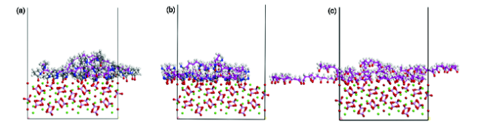
体系的平衡可由温度和能量同时到达平衡予以确定。通常当温度和能量在5%~10%范围内波动即可认为体系已达到平衡。本研究的所有计算都是在体系达到平衡的条件下进行的。经MD模拟, 得到了三种高分子材料与HA(110)的界面相互作用的平衡结构模型, 如图1所示。其中紫色为高聚物的碳主链, 灰色为体系中主链碳原子外的碳原子, 红色为原子氧, 乳白色为氢原子, 蓝色为氮原子, 紫色为磷原子, 绿色为钙原子。
图1 三种粘结剂与HA界面相互作用模型的平衡结构
Fig.1 Equilibrium structures of binder/HA interfacial interaction models. (a) PVP/HA(110) interaction model; (b) PAM/HA(110) interaction model; (c) PVA/HA(110) interaction model
真实的高分子链所含原子个数成千上万, 在模拟过程中构建一条真实的高分子链是不切实际的, 这是由计算能力的客观因素所决定的。因此, 高分子链聚合度的选取是模拟成功的关键, 高分子链所含单元体个数既不能超出计算量的上限, 还必须能够代表高分子的性质。内聚能密度(Cohesive Energy Density, CED)是表征高分子材料的一个重要参数, 是对物质分子间相互作用力大小的度量, 凡与物质间相互作用有关的性质和物性属性与内聚能密度都具有内在联系, 如物质的溶解性、相容性、汽化热等, 其表达式如下:
式中Ecoh是指内聚能,
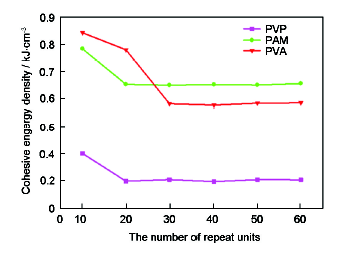
为了选取合适的高分子聚合度, 本文构建了含不同聚合度的PVP、PAM、PVA分子链模型, 并对其无定型晶胞进行了MD模拟, 计算得到了不同聚合度高分子链的内聚能密度, 图2为内聚能密度与高分子聚合度的关系。
通过内聚能密度与聚合度的关系图, 可见PVP、PAM的聚合度超过20后, 内聚能密度就趋于稳定, 而PVA则要超过30才能趋于稳定。而本文为了对三种材料进行对比, 同时权衡计算能力, 最终将PVP、PAM、PVA的聚合度取为20、31、50, 即单链的分子量相近。此外通过比较三种高聚物稳定时的CED, 其大小关系为PAM>PVA>PVP; 而CED的大小可以反映物质的稳定性以及其内部分子间相互作用的强弱, 即内聚能密度越大物质的稳定性、内部相互作用力也就越大, 所以高聚物体系的稳定性及其内部相互作用的强弱关系为PAM>PVA> PVP。
高聚物与HA(110)界面的结合能是高聚物分子与HA界面分子相互作用力大小的一个量化, 可以反映出高聚物粘结剂与HA表面的粘结作用力大小, 是粘结剂的一个重要参数。其表达式如下:
式中,
由表2数据可见, PVP、PAM、PVA三种粘结剂高分子材料在HA表面的结合能大小关系为PAM>PVA>PVP。说明这三种粘结剂与HA界面粘合力的大小关系为PAM>PVA>PVP, 即PAM对HA界面的粘结力相对最强。而组分间结合能的大小是界面分子相互作用力的反映, 结合能越大, 产生的分子间相互作用力越大, 这与内聚能密度的大小关系相一致。导致上述结果的最根本原因是因为相同质量的三种粘结剂所含有的官能基团的极性、数目不等, 1条PVP链含有20个羰基(-C=O), 1条PVA链含有50个羟基(-OH), 而1条PAM链则含有31个酰胺基(-NH2)、31个羰基(-C=O)。
表2 高聚物与HA(110)界面相互作用体系的结合能
Table 2 The binding energies between polymers and HA(110) surface (kJ/mol)
| System | Etotal | Eploymer | EHA | Ebind |
|---|---|---|---|---|
| PVP/HA | 398067331 | 36773 | 39917761 | 147803 |
| PAM/HA | 39706547 | 58660 | 39917652 | 269765 |
| PVA/HA | 39744011 | 49373 | 39917778 | 223140 |
为了进一步阐述粘结剂与HA界面间粘结机理及本质, 本文对三种粘结剂与HA界面相互作用体系的平衡结构进行对关联函数(Pair Correlation Function, PCF)分析。对关联函数g(r)可表示在距离某一指定参考原子A为r处, B原子出现的概率, 其表达式具体如下。
式中nB表示在距离A原子半径为r的范围内B原子的数目, NB表示整个体系中B原子的总数,
在进行对关联函数的分析之前, 首先必须对相互作用体系中的各类原子进行标记, PVP分子链中的原子类型有主链C原子、与主链C原子直接相连的H原子、 N原子、与N原子构成五元环的C原子以及羰基(-C=O)O原子, 其中只需对可能与周围原子发生较强相互作用的原子进行标记, 所以将与主链C原子直接相连的H原子、N原子、羰基(-C=O)O原子依次标记为H(PVP)、N(PVP)、O(-C=O); 类似的, 在PAM中将与主链C原子直接相连的H原子、羰基(-C=O)O原子、酰胺基(-NH2)N原子、酰胺基H原子标记为H(PAM)、O(-C=O)、N(-NH2)、H(-NH2); 在PVA分子链中, 将羟基(-OH)O原子、羟基H原子、与主链C直接相连的H原子依次标记为O(-OH)、H(-OH)、H(PVA); 将HA中的Ca原子、磷酸根(-PO3-4)中的O原子、H原子和羟基(-OH)中的O原子, 依次表示为Ca、O(HA1)、H(HA)、O(HA2); 本文分别对上述三种高聚物所标记的原子与HA中所标记的原子之间进行了对关联函数分析(图3-4), 此外还对三种粘结剂高分子C主链在HA界面的分布进行了分析(图3)。图4只给出了存在重要相互作用的g(r)~r关系。
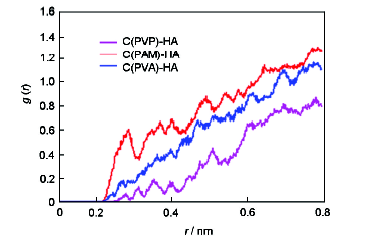
图3为三种粘结剂高分子C主链与HA界面的对关联函数, 由图可见PVP、PAM、PVA的C主链所对应的对关联函数值的大小关系为PAM>PVA>PVP, 而C主链可以在一定程度上代表整个高分子, 所以函数值的大小可以反映出粘结剂与HA界面粘合力的大小, 即相互作用力越大, 其在距离HA表面同一距离r处的函数值也越大。这一结论与粘结剂与HA相互作用结合能的大小关系是一致的, 进一步验证了计算结果的正确性。
图3 主链C原子与HA界面的对关联函数
Fig.3 The PCFs between the carbon backbone and HA interface
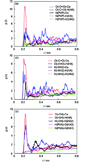
分子间相互作用力一般分为化学键、氢键作用和范德华作用, 对关联函数图中0.35 nm以内的峰值主要由化学键、氢键构成, 0.35 nm以外的则主要是库伦、范德华相互作用成分[18]。图4a分析了PVP/HA界面相互作用模型中存在重要相互作用的原子间对关联函数, PVP粘结剂中的羰基(-C=O)O原子、N原子与HA中Ca原子的对关联函数的峰值位于r=0.22 nm附近, 比Ca-O离子键键长0.239 nm、Ca-N离子键键长0.247 nm均小, 表明PVP中的羰基O原子、N原子分别与HA中的Ca原子之间形成了离子键; 此外, PVP中的羰基O原子、N原子与H(HA)原子对关联函数的峰值位于r=0.21 nm附近, 说明它们之间主要是通过氢键发生相互作用; 而H(PVP)与O(HA2)对关联函数的峰值不是特别明显, 但是也形成了少部分氢键, 大部分是范德华作用; 由上述原子间对关联函数的峰值大小, 可以发现PVP与HA界面相互作用主要是靠PVP中的极性原子、官能团与HA界面原子形成离子键、氢键, 且离子键的作用强度最大。
图4 粘结剂/HA界面相互作用的对关联函数
Fig.4 PCFS of binder/HA interface interaction: (a) PCFS of PVP/HA interface interaction; (b) PCFS of PAM/HA interface interaction; (c) PCFS of PVA/HA interface interaction
图4b为PAM/HA界面相互作用的对关联函数, 由图可见, PAM中的羰基O原子、酰胺基N原子与HA中Ca原子间的对关联函数峰值分布在r=0.22 nm附近, 说明它们之间形成了离子键; 此外, 羰基O原子、酰胺基N原子与HA中羟基H的对关联函数峰值也在氢键的作用范围之内, 说明它们之间形成了氢键作用; 而酰胺基H原子与HA中羟基O原子间的相互作用不是很强, 主要是发生范德华作用; 从上述原子间对关联函数的峰值大小分析可以看出PAM与HA界面相互作用主要是通过羰基O原子、酰胺基N原子与HA界面原子形成离子键、氢键, 且离子键作用强度最大, 其次是氢键作用。
图4c为PVA/HA界面相互作用的对关联函数, 由图可见PVA中羟基(-OH)O原子与HA中的Ca原子、羟基H原子间的对关联函数峰值分别处于r=0.21~0.22 nm附近, 说明其分别形成了离子键、氢键作用; 而PVA中羟基H原子与HA中羟基O原子间的对关联函数的峰值位于r=0.31 nm附近, 这是氢键的作用范围, 说明它们之间是通过氢键发生相互作用; PVA中与主链C原子直接相连的H原子与HA中O原子间的相互作用很微弱, 主要是范德华作用; 比较对关联函数峰值的大小可以发现PVA与HA界面相互作用也主要是通过PVA中的羟基与HA界面原子形成离子键、氢键, 且离子键作用强度最大, 氢键次之。
所以通过对PVP、PAM、PVA三种粘结剂高分子与HA的对关联函数图像分析, 可以发现, 粘结剂与HA发生相互作用主要是通过高聚物中的极性官能基团与HA中的Ca原子、羟基发生作用形成离子键、氢键, 且离子键作用强度最大, 氢键次之。
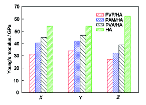
骨支架的制备过程中, 力学性能是选用粘结剂的一个重要指标, 本文应用Materials Studio 软件中的Forcite模块, 对三种粘结剂与HA相互作用平衡结构X、Y、Z三个方向(Z为作用面的垂直方向)的杨氏模量进行了MD模拟计算, 其计算结果如图5所示。
由图5不难发现三种粘结剂与HA相互作用模型三个方向的杨氏模量并不一致, 存在一定差异, 这一结论与HA的各向异性相符, 即粘结剂的添加并没有改变材料的各向异性; 添加粘结剂后的相互作用模型, 其各个方向的力学性能较单一HA力学性能均有所下降, 表现最为明显的是Z方向的力学性能, 这主要与建立相互作用模型时作用面的选取有关, 即本文是在HA超级晶胞Z方向添加粘结剂; 比较三个相互作用模型的力学性能, 可以发现其力学性能存在一定的优劣关系, 力学性能最好的是PVA/HA, 其次是PAM/HA, 最低的是PVP/HA, 即PVA>PAM>PVP, 这一结论与结合能的大小关系并不完全一致, 这说明相互作用模型的力学性能与粘结剂的粘性并不存在特定的内在关系。
1. 通过计算比较三种粘结剂稳定时的内聚能密度, 得出了三种材料的稳定性及其内部相互作用的强弱关系为PAM>PVA>PVP。
2. 结合能的大小是界面分子相互作用力大小的反映, 结合能越大, 分子间相互作用力就越大, 所以由结合能的大小关系得出了三种粘结剂与HA界面粘合力的大小关系, 即为PAM>PVA>PVP, 这与内聚能密度的大小关系相一致。
3. 通过对PVP、PAM、PVA三种粘结剂高分子与HA的对关联函数分析, 可以发现, 粘结剂与HA发生相互作用主要是通过高聚物中的极性官能基团与HA中的Ca原子、羟基发生作用形成离子键、氢键, 且离子键作用强度最大, 氢键次之。
4. 三种相互作用模型各个方向的力学性能较单一HA有所降低, 且Z方向尤为明显。对比三个相互作用模型的力学性能, 其粘结模型力学性能的优劣关系为PVA>PAM>PVP, 这一结论与结合能的大小关系并不完全一致, 这说明相互作用模型的力学性能与粘结剂的粘性并不存在特定的内在关系。
The authors have declared that no competing interests exist.
| [1] |
Progress in the biomineralization Study of bone and enamel and biomimetic synthesis of Calcium phosphate, 骨与牙釉质组织的生物矿化及磷酸钙材料仿生合成研究进展,
<FONT face=Verdana>骨和牙釉是典型的有机基质介导生成的生物矿化材料,其中的矿物相都属于以磷灰石为主的钙磷酸盐系统,但有机基质的不同使得晶体尺寸、形貌及排列方式迥异.本文综述了有关骨和牙釉组织的生物矿化研究,重点探讨对这些天然生物矿化组织的分级结构、基质蛋白的自组装及调控矿化机理的认识.在此基础上,磷酸钙材料的仿生合成以期应用于硬组织的缺损修复或再生医学也是目前的重要研究内容.</FONT>
|
| [2] |
Bone regeneration in osseous defects using hydroxyapatite graft and the extent of ossification in osseous defects treated without graft: a comparative evaluation ,
Objective of this study is to evaluate the new bone formation in bony defects following insertion of hydroxyapatite graft and to compare the efficacy and regenerative potential of this bone graft material.The patients having osseous defects after surgery were selected. Preoperatively a brief history, examination, relevant blood investigation and radiographs were taken. Post operative observation period of 6months was planned. Radiographic and bone scintigraphic (isotope study of bone activity) evaluation of bone specimens was completed in defined time.Radiographic evaluation indicated increased calcification surrounding the material, indicative of acceptance of the graft to the bone. Bone scintigraphic evaluation indicated area of increased bone metabolism and is evidenced as area of increased radiotracer uptake, namely 'hot spots' (active bone formation).It can be concluded that the hydroxyapatite graft met the clinical requirement of a bone substitute material which is biocompatible and non-allergic. The use of this material is advantageous over other bone grafts because of simplicity of application, cost effectiveness and easy availability. Due to its microstructure, complete resorption and neo bone formation took place during the course of this study.
|
| [3] |
Preparation, characterization and antimicrobial activity of a bio-comosite scaffold containing chitosan/nano-hydroxyapatite/nano-silver for bone tissue engineering, Int. J .
In this study, a bio-composite scaffold containing chitosan/nano-hydroxyapatite/nano-silver particles (CS/nHAp/nAg) was developed by freeze drying technique, followed by introduction of silver ions in controlled amount through reduction phenomenon by functional groups of chitosan. The scaffolds were characterized using SEM, FT-IR, XRD, swelling, and biodegradation studies. The testing of the prepared scaffolds with Gram-positive and Gram-negative bacterial strains showed antibacterial activity. The scaffold materials were also found to be non-toxic to rat osteoprogenitor cells and human osteosarcoma cell line. Thus, these results suggested that CS/nHAp/nAg bio-composite scaffolds have the potential in controlling implant associated bacterial infection during reconstructive surgery of bone.
|
| [4] |
Composite chitosan/ nano-hydroxypatite scaffolds induce osteocalcin production by osteoblasts in vitro and support bone formation in vivo, Tissue Eng.
There is a significant clinical need to develop alternatives to autografts and allografts for bone grafting procedures. Porous, biodegradable scaffolds based on the biopolymer chitosan have been investigated as bone graft substitutes, and the addition of calcium phosphate to these scaffolds has been shown to improve the mechanical properties of the scaffold and may increase osteoconductivity. In this study, in vitro mineralization was examined for osteoblasts seeded in a porous scaffold composed of fused chitosan/nano-hydroxyapatite microspheres. Human fetal osteoblasts were cultured on composite and chitosan scaffolds for 21 days. On days 1, 4, 7, 14, and 21, total dsDNA, alkaline phosphatase, type I collagen, and osteocalcin production were measured. Total cellularity (measured by dsDNA), alkaline phosphatase, and type I collagen production were similar between the two scaffold groups. However, osteocalcin production occurred significantly earlier (day 7 vs. day 21) and was more than three times greater (0.0022 vs. 0.0068 ng/mL/ng DNA) on day 21 when osteoblasts were cultured on composite scaffolds. Osteocalcin is a marker of late osteoblastic differentiation and mineralized bone matrix formation. Therefore, the increase in osteocalcin production seen when cells were cultured on composite scaffolds may indicate that these scaffolds were superior to chitosan-only scaffolds in facilitating osteoblast mineralization. Composite scaffolds were also shown to be biocompatible and osteoconductive in a preliminary critical size rat calvarial defect study. These results demonstrate the potential of composite chitosan/nano-hydroxyapatite scaffolds to be used in bone tissue engineering.
|
| [5] |
Direct scaffolding of biomimetic hydroxyapatite-gelatin nanocomposites using aminosilane cross-linker for bone regeneration, J .
Hydroxyapatite-gelatin modified siloxane (GEMOSIL) nanocomposite was developed by coating, kneading and hardening processes to provide formable scaffolding for alloplastic graft applications. The pres
|
| [6] |
Three dimensional printing: Rapid tooling and prototypes directly from a CAD model, |
| [7] |
Review of research status for Three-Dimensional printing technology in rapid prototyping of powder material, 三维打印技术在粉末材料快速成形中的研究现状评述,
自20世纪80年代中后期基于分层制造思想的快速成形(RP)技术出现以来,零件或模具的直接快速精密制造已成为加工制造业的重要发展方向之一;而随着粉体制备技术的不断提高,许多粉末材料已商品化,成本也逐渐降低,采用粉末材料直接成形零件或模具已成为当前的研究热点.目前,用于粉末材料快速成形的主要工艺有基于激光技术的选择性激光烧结(SLS)和基于微喷射粘结技术的三维打印(3DP).
|
| [8] |
In vitro bioactivity of bioresorbable porous polymeric scaffolds incorporating hydroxyapatite microspheres, DOI URL PMID Magsci [本文引用: 1] 摘要
<h2 class="secHeading" id="section_abstract">Abstract</h2><p id="">Biomimetic composites consisting of polymer and mineral components, resembling bone in structure and composition, were produced using a rapid prototyping technique for bone tissue engineering applications. Solid freeform fabrication, known as rapid prototyping (RP) technology, allows scaffolds to be designed with pre-defined and controlled external and internal architecture. Using the indirect RP technique, a three-component scaffold with a woodpile structure, consisting of poly-<span class="smallcaps">l</span>-lactic acid (PLLA), chitosan and hydroxyapatite (HA) microspheres, was produced that had a macroporosity of more than 50% together with micropores induced by lyophilization. X-ray diffraction analysis indicated that the preparation and construction of the composite scaffold did not affect the phase composition of the HA. The compressive strength and elastic modulus (<em>E</em>) for the PLLA composites are 0.42 and 1.46 MPa, respectively, which are much higher than those of chitosan/HA composites and resemble the properties of cellular structure. These scaffolds showed excellent biocompatibility and ability for three-dimensional tissue growth of MC3T3-E1 pre-osteoblastic cells. The pre-osteoblastic cells cultured on these scaffolds formed a network on the HA microspheres and proliferated not only in the macropore channels but also in the micropores, as seen from the histological analysis and electron microscopy. The proliferating cells formed an extracellular matrix network and also differentiated into mature osteoblasts, as indicated by alkaline phosphatase enzyme activity. The properties of these scaffolds indicate that they can be used for non-load-bearing applications.</p>
|
| [9] |
Effects of pore size and void fraction on cellular adhesion, proliferation, and matrix deposition ,
The aim of this study was to determine the influence of two key scaffold design parameters, void fraction (VF) and pore size, on the attachment, growth, and extracellular matrix deposition by several cell types. Disc-shaped, porous, poly(-lactic acid) (L-PLA) scaffolds were manufactured by the TheriForm solid free-form fabrication process to generate scaffolds with two VF (75% and 90%) and four pore size distributions (38 to 150 microm; however, when cultured on scaffolds with pores formed with salt particles of <38 microm, MVEC formed a multilayered lining on the scaffolds surface. Culture data from scaffolds with a 75% VF suggests that the structural features were unsuitable for tissue formation. Hence, there were limits of acceptable scaffold architecture (VF, pore size) that modulated in vitro cellular responses.
|
| [10] |
Eng.
|
| [11] |
Processing of polycaprolactone porous structure for scaffold development,
The study investigated the processing of biodegradable porous scaffold of polycaprolactone (PCL). Commercially available PCL powder was blended with poly(vinyl alcohol) powder at various compositions to fabricate pre-scaffolds using the three-dimensional printing (3DP) technique followed by particulate-leaching. Structures at different porosity (50–70%) were processed and their properties were characterised using differential scanning calorimetry, dynamic mechanical analyser, scanning electronic micrography and gel-permeable chromotography. The scaffolds produced in this route displayed open cellular structures with macropores with pores sizes ranging between 100 and 500 渭m. No significant degradation of PCL was detected. The storage modulus of the scaffold ranged 0.03–0.19 MPa.
|
| [12] |
Inkjet printing of highly loaded particulate suspensions,
ABSTRACT Inkjet printing is an attractive method for patterning and fabricating objects directly from design or image files without the need for masks, patterns, or dies. In order to achieve this with metals or ceramics, it is often necessary to print them as highly concentrated suspensions of powders in liquids. Such liquid suspensions must have physical properties appropriate to the inkjet delivery mechanism. These properties are presented using a nondimensional formalism to illustrate the requirements for both drop formation and spreading on impact. Further critical issues relevant to inkjet printing of particulate suspensions are discussed and illustrated with experiments on a model alumina-containing colloidal suspension.
|
| [13] |
Crystal structure of hydroxylapatite, |
| [14] |
羟基磷灰石/α-氰基丙烯酸正丁酯相互作用及力学性能的MD模拟,
基于羟基磷灰石陶瓷微球人工骨支架的制备工艺,用分子动力学(MD)方法研究了羟基磷灰石(HA)的三个晶面(001)、(100)、(110)分别与生物胶黏剂α-氰基丙烯酸正丁酯(NBCA)相互作用后混合体系的结合能,并计算分析了(110)晶面的力学性能和径向分布函数。结果表明,三晶面所对应结合能关系为HA(110)HA(100)HA(001);HA(110)晶面与NBCA之间粘结强度高于HA(100)和HA(001)晶面;对聚合度为40的NBCA聚合物与HA相互作用的MD计算表明,HA(110)/NBCA混合体系的力学性能比单组份HA体系有所下降,但满足人工骨对力学性能的要求;元素之间的径向分布函数揭示了混合体系HA/NBCA组分之间的相互作用机理,NBCA与HA(110)晶面存在较强的相互作用。其主要原因是,NBCA中的N原子和羰基中的O原子分别与HA中的H原子形成的氢键,进而说明了HA对NBCA的强吸附作用。
|
| [15] |
Molecular dynamics simulations at constant pressure and /or temperature, |
| [16] |
Cohesion of ionic solids in the Born model,
Data on the postembryonic development, longevity, ageing and rejuvenation of planarians are reviewed. From the available information, including the results of their own experiments, the authors conclude that planarians display a pattern of ageing intermediate between that of the "non-ageing" (fully-renewable) invertebrates and typical senescence as seen in mammals. The reversibility of planarian age changes and the measurement of "rejuvenation" in planarians are discussed.
|
| [17] |
Evaluation of optical and electrostatic lattice potentials, |
| [18] |
Micro theoretical study on the compatibility and mechanical properties of polyacrylamide/polyvinyl alcohol blends, 聚丙烯酰胺/聚乙烯醇共混物相容性及力学性能的微观理论研究,
采用分子动力学的理论方法,从微观分子相互作用的层面研究了 PAM/PVA 共混体系,分别从组分间的相容性、结合能以及对相关函数分析3方面阐述了共混体系间相互作用的机理及本质,通过对组分不同质量配比体系的静态力学分析,研究了组分不同质量配比对体系力学性能的影响。结果表明,PAM 与PVA 组成的共混物相容性极好,而体系中单组分间的结合能随着 PVA 含量的增加反而减小,主要原因是质量相同的 PAM 链所含极性官能团的数目要高于 PVA链;此外,对相关函数分析表明,单组分间主要是通过氢键形成相互作用,且共混体系中酰胺基(—NH 2)、羰基(—C ??O)、羟基(—OH)官能团与周围 H 原子形成氢键的可能性关系为 O (-C=O)> O (-OH)>N(-NH2),这也是酰胺基(—NH 2)、羰基(—C ?? O )、羟基(—OH)官能团极性强弱关系的反映;单组分不同质量配比模型的静态力学分析表明,随着共混体系中 PVA组分含量的增加,体系弹性系数、各项工程模量、柯西压值均呈上升趋势,即 PVA 含量的增加可以明显改善PAM 的力学性能及其延展性。
|

/
| 〈 |
|
〉 |