
李晨曦 , 任婧
, 任婧
LI Chenxi, REN Jing
文献标识码: 分类号 R783.1 文章编号 1005-3093(2016)07-0489-07
通讯作者:
收稿日期: 2015-09-7
网络出版日期: 2016-07-25
版权声明: 2016 《材料研究学报》编辑部 《材料研究学报》编辑部
作者简介:
本文联系人: 梁锐英
展开
摘要
以高度抛光的玻璃陶瓷、聚合瓷、氧化锆、纯钛为摩擦副, 模拟口腔环境, 使用微摩擦磨损实验机进行天然牙釉质和牙本质的摩擦磨损实验。用扫描电镜观察表面形貌、用粗糙度仪测粗糙度、用维氏硬度仪测表面硬度、用电子天平测磨损量, 研究了天然牙釉质和牙本质与不同修复材料之间的摩擦磨损性能。结果表明, 釉质和牙本质分别与四种材料对磨后的磨损量与对照组均有统计学差异(P<0. 05), 对磨物的磨损量与四种材料及釉质、牙本质的硬度值呈显著正相关关系。其中牙本质与聚合瓷对磨后的磨损量与牙本质对照组最接近, 釉质与玻璃陶瓷对磨后的磨损量与釉质对照组最接近。
关键词:
Abstract
The friction and wear behavior of the natural tooth enamel and dentin in sliding against a highly polished glass ceramic, polymer ceramic, zirconia and pure titanium respectively were investigated through a fretting friction and wear testing in an artificial saliva test environment by UMT-2 friction and wear testing machine. While the worn surface morphology was observed by scanning electron microscopy (SEM), the roughness was measured by roughness instrument and the abrasion loss was weighed by an electronic balance. The results show that after friction and wear testing, the abrasion loss of the enamel and dentin against the four kind of materials all showed a statistically difference within P < 0. 05, which possessed significant positive correlation with the hardness of the four kinds restoration materials, the enamel and the dentin. The mass loss of the dentin against poly ceramic and the enamel against glass ceramic was the closest ones to that of the control group of the dentin.
Keywords:
随着口腔材料学的发展, 越来越多性能优良的修复材料应用于临床。理想的口腔修复材料的硬度、色泽和通透性应该与天然牙相似, 尤其是其磨损性能。在咀嚼过程中, 冠修复体会使自身及对颌天然牙产生一定程度的磨耗, 过度磨损牙体组织造成釉质脱落、牙本质过敏、牙髓炎和颞颌关节紊乱等疾病, 过度磨损冠修复体则会缩短其使用寿命。目前临床常用的冠修复材料有金属、陶瓷、树脂等材料[1]。口腔咀嚼食物发生摩擦运动时咬合力、温度、唾液、pH等因素的变化, 均对修复材料和天然牙的耐磨性产生重大影响。材料的物理性能、修复体的使用时间等与磨耗程度也有密切的关系。釉质、牙本质的硬度和结构的差异使同种修复材料对釉质和牙本质造成不同的磨耗, 不同修复材料对牙体组织产生的磨耗程度也不同。以往的研究多用滑石瓷替代釉质作为实验对照, 且材料只与釉质对磨, 与临床情况有一定的不同。本文以天然牙釉质和牙本质牙尖为研究对象, 模拟口腔环境, 选取纯钛、氧化锆、玻璃陶瓷、聚合瓷四种材料与天然牙对磨, 研究其摩擦磨损特征。
材料: 纯钛金属(BEGO), 氧化锆, 玻璃陶瓷(VITA), 聚合瓷, 环氧树脂, 两周内拔除的天然牙(表面无磨耗, 无龋坏, 无脱矿现象), 应用ISO/ TR 10271 标准制备人工唾液(成分: NaCl: 0.4 g; KCl: 0.4 g; CaCl22H2O: 0.795 g; NaH2PO4H2O2: 0.78 g; Na2S9H2O: 0.005 g; Urea: 1 g; Distilled Water: 1000 g)[2]。
设备: 微摩擦实验机(UMT-2, CETR, USA), 粗糙度仪(MarSurf M 300 C + RD 18 C, Mahr GmbH ), HVS-50Z型自动转塔数显维氏硬度仪, 扫描电子显微镜(S-4800), 抛光用金刚砂磨头(DFS), 电子天平, 超声振荡清洗器。
选取因正畸拔除的未磨耗双尖牙或未萌出的第三磨牙, 拔除后立即放入生理盐水中并保存在4℃的冰箱中[3]。磨取釉质和牙本质牙尖各50个作为实验上试件, 釉质牙尖包埋于红色的环氧树脂, 牙本质包埋于白色环氧树脂中。
按照实验机的要求将上试件制作成直径3.1 mm, 高12 mm的销形试样[3], 对磨牙尖接触端为至少0.5 mm小平面, 齿科专用抛光轮由粗到细依次抛光, 超声震荡清洗后, 保存于生理盐水中。
下试件实验组包括纯钛, 氧化锆, 玻璃陶瓷, 聚合瓷各20个, 规格为15×10×5 mm的平面试件; 对照组包括片切天然牙颊侧釉质及牙本质各10个, 厚度不小于2 mm, 包埋于环氧树脂中, 规格与实验组的相同, 暴露天然牙至少5×5 mm平面。将所有下试件用200-600目碳化硅砂纸依次打磨后, 分别用专用抛光磨头抛光至最小粒度, 超声清洗后备用[3]。
将釉质及本质牙尖随机分为5组(n=10), 用不同颜色指甲油标记为ABCDE, 从每组随机抽取一个试件置于同一个容器中(共十个容器1-10)。将同种材料的下试件随机分为2组(n=10), 用不同颜色指甲油标记为釉质组、本质组, 依次在每组试件侧面用高速手机标记1-10, 对照组的标记与实验组的相同。
1.3.1 硬度的测量 用HVS-50Z型自动转塔数显维氏硬度仪测量釉质、牙本质、玻璃陶瓷、聚合瓷、氧化锆、纯钛的表面显微硬度。负荷为98 N, 时间为15sec, 在每种材料的表面选10个点测量, 取10个测量值的平均值作为该材料的硬度值。
1.3.2 粗糙度的检测 用MarSurf 粗糙度仪测量釉质、牙本质、玻璃陶瓷、聚合瓷、氧化锆、纯钛的表面粗糙度。扫描长度Lt=4.0 mm, 截止波长0.800 mm, 取样长度n=5。在每个试件抛光面的中心区域测十次, 取其平均值为该试件的表面粗糙度, 再取十个试件的粗糙度平均值作为该材料的粗糙度。
1.3.3 摩擦实验 使用微摩擦实验机(UMT-2, CETR, USA)进行销盘式往复摩擦实验[4], 测试天然牙釉质、牙本质分别与玻璃陶瓷、聚合瓷、氧化锆、纯钛的摩擦磨损性能。有无人工唾液介质对摩擦实验的结果影响很大, 因此在实验中用人工唾液浸润。正常人咀嚼时上下对颌牙的滑动距离约为0.9-1.2 mm[5], 本文设计垂直载荷为15 N, 摩擦运动方式为往复运动, 位移幅度1 mm, 频率2 Hz, 循环次数6000次。用计算机控制并记录实验过程中表面摩擦力与位移在不同循环次数下的变化, 以得到摩擦系数(cof)等参数的变化曲线[6]。
1.3.4 质量损失 将所有上试件置于蒸馏水中清洗10 min, 用无水乙醇和丙酮交替冲洗三次, 用冷风吹干后用电子天平测量质量(精确至0.001 mg), 实验后用相同程序称质量, 计算质量损失量。
1.3.5 电镜扫描表面形貌 用扫描电镜观测釉质、牙本质磨损实验前后牙尖表面磨斑的微观形貌。
1.3.6 统计学分析 使用SPSS17.0软件包分析实验数据, 对材料粗糙度、硬度及对磨牙尖质量损失量进行单因素方差分析和两两比较。分析材料引起的釉质、牙本质牙尖磨损量与硬度之间的关联性。检验水准α= 0. 05。
测量结果如图1所示, 图中所示为各组粗糙度均值, 可见实验组的粗糙度均显著高于对照组, 对各实验组粗糙度的单因素方差分析及两两比较结果表明, P>0.05, 没有统计学差异, 材料表面的粗糙度没有差异。
由图2可见, 试件的硬度由小到大依次为牙本质、聚合瓷、纯钛、釉质、玻璃陶瓷、氧化锆。聚合瓷与牙本质的硬度最接近, 纯钛的比釉质略低, 玻璃陶瓷的略高于釉质, 氧化锆的显著高于其他材料。材料的硬度表示其抗弹性形变的能力, 是耐磨性的重要影响因素。硬度与材料磨损量之间呈一定的正相关关系, 硬度越高的材料越耐磨, 因此修复材料应该有与天然釉质及牙本质相近的硬度 [7]。
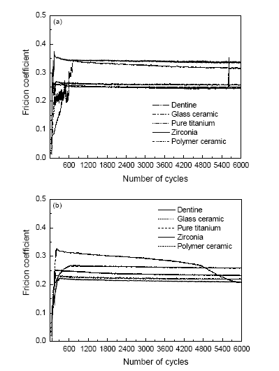
2.2.1 摩擦系数 摩擦系数(cof)是指两表面间的摩擦力与作用在其一表面上的垂直力之比值[8]。基于人体口腔生理环境的特性, 口腔修复材料与天然牙组成磨擦副, 耐磨性是否匹配对摩擦副的正常行使功能有重要的意义。口腔修复材料应具有与天然牙尽可能接近的磨耗性能, 即耐磨性低于天然牙, 但又与其接近[9]。cof作为反映材料摩擦学性能的一项参数, 其值越小表明摩擦性能越好, 已知cof与材料接触面粗糙度有关, 指导临床调磨修复体后应进行高度抛光, 降低其cof, 减少对颌牙及修复体的磨损[10]。本文的实验模拟口腔环境在常温人工唾液浸泡下进行摩擦实验, 所得摩擦系数如图3所示。
图3 本质各组(a)和釉质各组(b)的摩擦系数
Fig.3 Friction coefficient of dentine (a) and enamel (b)
2.2.2 质量损失 表1给出了釉质、牙本质牙尖与不同材料对磨后的质量损失量。可见牙本质组中牙尖的质量损失由大到小依次为氧化锆组、纯钛组、玻璃陶瓷组、聚合瓷组、牙本质组, 其中聚合瓷组与牙本质组最相近; 釉质组中牙尖的质量损失由大到小依次为氧化锆组、玻璃陶瓷组、釉质组、纯钛组、聚合瓷组, 其中玻璃陶瓷组与釉质组最接近, 纯钛组略低于釉质组。
表1 牙尖质量损失量
Table1 Wear loss of tooth cusp (mg)
| Group | Wear loss of dentine cusp (mg) | Group | Wear loss of enamel cusp (mg) |
|---|---|---|---|
| Dentine | 0.364±0.0171270 | Enamel | 0.308±0.0690893 |
| Glass ceramic | 0.490±0.0447214 | Glass ceramic | 0.320±0.0339935 |
| Polymer ceramic | 0.368±0.0293636 | Polymer ceramic | 0.170±0.0258199 |
| Zirconia | 0.812±0.0418463 | Zirconia | 0.562±0.0367575 |
| Pure titanium | 0.720±0.0258199 | Pure titanium | 0.272±0.0355278 |
表2给出了釉质、牙本质及四种材料的硬度、粗糙度。使用SPSS17.0软件包对实验组粗糙度进行单因素方差分析及两两比较[11], 结果表明P>0.05, 四种材料间没有统计学差异。对牙本质、釉质及四种材料的硬度进行单因素方差分析及两两比较, 表明P<0.05, 材料的硬度不完全相同。对牙本质组的牙尖质量损失量进行单因素方差分析, 表明P<0.05, 差异具有统计学意义, 即五组材料间的质量损失量不完全相同, 两两比较可得牙本质和聚合瓷组之间的p值>0.05, 即两组间的质量损失差异不具有统计学意义。对釉质组进行单因素方差分析, 表明P<0.05, 五组材料间的质量损失量不完全相同, 两两比较结果牙釉质和纯钛组、牙釉质和玻璃陶瓷组之间的p值>0.05, 即其差异没有统计学意义。
表2 材料硬度及粗糙度
Table 2 Hardness and roughness of materials
| Group | Hardness of materials (HV) | Roughness of materials (Ra) |
|---|---|---|
| Dentine | 54.11±0.8293 | 0.17770±0.026874 |
| Enamel | 609.52±9.3542 | 0.27490±0.013536 |
| Glass ceramic | 609.52±9.3542 | 0.27490±0.013536 |
| Polymer ceramic | 64.09±0.4557 | 0.27880±0.024068 |
| Zirconia | 1311.83±39.9536 | 0.28170±0.033859 |
| Pure titanium | 239.72±3.4071 | 0.27420±0.016772 |
研究结果表明, 材料的硬度越大对天然牙的磨损越严重[12], 因此对四种材料引起的对磨物的质量损失量与材料硬度之间进行关联性分析。牙本质组r=0.746, 为中度相关, P=0.147, >0.05, 表明相关不显著, 硬度与质量损失之间有一定的正向趋势。釉质组r=0.979, >0.8, P=0.004, <0.05, 表明相关性显著, 可见硬度与质量损失之间有高度的正相关性, 即随着硬度的增加质量损失量随之增加。
2.2.3 磨损形貌 图4(A-E)给出了牙本质与各组材料对磨前后的表面微观形貌; 图5(a-e)给出了釉质与各组材料对磨前后的表面微观形貌。图4A表面有明显的塑性变形, 有沟槽状划痕, 提示疲劳磨损为其主要磨损类型, 伴有一定程度的磨粒磨损。图4C表面磨斑比较光滑, 呈现出龟裂和组织的片状剥离, 提示疲劳磨损为其主要的磨损类型。图4BDE表面的磨斑清晰, 呈现明显的犁沟和碎屑。在犁沟间可见裂纹和片状剥脱, 表明磨粒磨损是其主要的磨损形式, 还伴有疲劳磨损[13]。图5a表明牙釉质局部区域有明显的塑性变形, 显微形貌呈现大量疲劳裂纹, 裂纹扩展, 生成片状剥离, 并有沟槽状划痕, 脱落的颗粒发生了一定的塑性变形, 表明磨损主要为疲劳磨损, 同时伴有黏着磨损和磨粒磨损[14]。图5b表明表面发生了片状剥离, 有些区域发生塑性变形后, 出现断裂, 提示磨损机制主要为疲劳磨损和磨粒磨损。图5c显示表面只有轻微的划痕和少量剥脱颗粒, 磨损面比较光滑, 主要发生疲劳磨损。图5d显示表面出现大量犁沟, 内附着大量剥脱颗粒, 提示主要发生磨粒磨损。图5e显示表面磨损出现明显的沟槽状划痕和剥脱碎屑, 提示发生了磨粒磨损和黏着磨损[14-15]。
图4 牙本质与各组材料对磨前后的表面微观形貌
Fig.4 Wear morphology of materials against dentine tip before and after experiment. (A) dentine cusp of control group, (B) dentine cusp against glass ceramic, (C) dentine cusp against polymer ceramic, (D) dentine cusp against zirconia, (E) dentine cusp against pure titanium
图5 釉质与各组材料对磨前后的表面微观形貌
Fig.5 Wear morphology of materials against enamel tip before and after experiment. (a) enamel cusp of control group, (b) enamel cusp against glass ceramic, (c) enamel cusp against polymer ceramic, (d) enamel cuspagainst zirconia, (e) enamel cusp against pure titanium
对于口腔材料, 工作条件就是人体口腔环境。实验结果表明, 在高应力作用下长时间摩擦磨损运动后, 在釉质及牙本质表面产生疲劳裂纹并扩展, 裂纹上的微粒剥落下来, 在表面形成深浅不同、大小不一的磨斑状凹坑, 脱落下来的微粒可重新压入材料表面, 表面所受的力传至表面下引起表面下分子键的断裂, 发生表面下的微破坏区, 引起表面破坏, 使物体表面发生的磨损为疲劳磨损; 也可在两界面间形成磨损颗粒, 引起表面擦伤, 形成犁沟, 即发生磨粒磨损。当两界面之间有较大的黏着力时, 在运动过程中一种物质表面发生破损, 破损碎屑融合到对方的表面, 即为黏着磨损。由此可见, 在材料与天然牙的磨损过程中几种磨损形式同时存在, 而且一种磨损诱发其他形式的磨损, 或以一种磨损形式为主伴随其他形式的磨损[16]。摩擦学理论表明, 材料的耐磨损性能不是材料的固有属性, 而是与磨损过程的工作条件密切相关的系统性能[17]。评价与天然牙相匹配的口腔材料, 其磨物磨损量应与釉质及牙本质相接近[18]。
在37℃人工唾液中进行实验, 釉质和牙本质分别与四种材料对磨后的磨损量与对照组均有统计学差异(P<0. 05), 对磨物的磨损量与玻璃陶瓷、聚合瓷、氧化锆、纯钛四种材料及釉质、牙本质的硬度值呈显著正相关关系。其中牙本质与聚合瓷对磨后的磨损量与牙本质对照组最接近, 釉质与玻璃陶瓷对磨后的磨损量与釉质对照组最接近。聚合瓷显示出类似于瓷的一些独特的特性, 其硬度和磨耗性能与牙本质相似。玻璃陶瓷的硬度及磨耗性能与釉质相似。根据对材料硬度及质量损失量之间的关联性分析, 应该临床选取与牙体组织硬度相近或略低的材料作为修复体。
致谢: 感谢清华大学摩擦学国家重点实验室给予的了大力帮助和支持。
The authors have declared that no competing interests exist.
| [1] |
LI Guo qiang, Wear resistance and hardness of dental prosthetics materials versus native teeth, 齿科修复材料耐磨性及硬度与天然牙齿的比较,
背景:天然牙与牙科修复材料之间存在摩擦现象,为了防止天然牙过 度磨损,同时延长修复体使用寿命,有必要了解天然牙与牙科修复材料间的摩擦学特性,选取相匹配的修复材料.目的:比较天然牙和各种牙科修复材料的摩擦学性 能及硬度.方法:由第一作者检索2000/2010 PubMed数据(http://www.ncbi.nlm.nih.gov/PubMed)及万方数据库(http://www. wanfangdata.com.cn)有关天然牙的磨耗以及各种牙科修复材料磨耗等方面的文献,英文检索词为"enamel,dental restorative material,zirconia, wear resistance,hardness",中文检索词为"牙釉质,口腔修复材料,氧化锆,耐磨性,硬度".计算机初检得到46篇文献,根据纳入标准保留 30篇进一步归纳总结. 结果与结论:牙釉质具有非常优良的摩擦学性能,耐磨性好,牙本质的耐磨性较差,天然牙齿在磨损过程中,牙本质一旦暴露,磨损将会加快.因此,选择耐磨性与 天然牙齿相匹配的牙科修复材料至关重要.牙釉质的耐磨性明显高于复合树脂,复合树脂材料对天然牙的磨损较小,但自身耐磨性较差.随着新型树脂材料物理性能 的不断提高,有些树脂材料的耐磨性已接近牙釉质.拜尔牙、热固塑料、铜基合金、钛及钛合金是较为理想的牙科修复材料.氧化锆陶瓷属于生物惰性陶瓷,具有很 好的生物相容性、高强度和韧性等特点,是一种新型的牙科修复材料.
|
| [2] |
Friction and wear properties of enamel opposing to dental materials used in dentistry, 六种常用冠修复材料的摩擦磨耗性能比较,
目的 探讨6种常用冠修复材料与滑石瓷对磨时的摩擦磨耗性能,为口腔常用冠修复材料的摩擦磨耗性能与牙釉质的匹配性提供依据.方法 使用MMJ-5G微机控制高温端面磨损实验机,以滑石瓷为对磨物,以15颗因正畸拔除的上颌第一前磨牙制作的10个试件为对照组,对纯钛(A组)、钴铬合 金(B组)、Superporcelain Ti-22体瓷(C组)、喜美乐体瓷(D组)、松风饰瓷(E组)、e.max饰瓷(F组)(每组均为10个试件)与滑石瓷组成的摩擦副在37℃人工唾液润 滑工况下进行摩擦磨耗实验.记录动态摩擦系数曲线、计算体积磨损百分比,扫描电镜观察表面磨损形貌.结果 试件体积磨损百分比由大至小为:对照组[(90.17×10⁻⁴)%]、A组[(79.23×10⁻⁴)%]、C组[(23.31 ×10⁻⁴)%]、D组[(20.41×10⁻⁴)%]、F组[(19.22 × 10⁻⁴)%]、E组[(8.53 × 10⁻⁴)%]、B组[(2.54×10⁻⁴)%].滑动摩擦系数由大到小依次是:D组(0.65)、C组(0.45)、E组(0.40)、A组 (0.35)、B组和F组(0.30).扫描电镜观察显示,E组表面最光滑,以下依次是F组、D组、C组.结论 纯钛与天然牙磨耗性能相近,是理想的修复金属材料.4种瓷粉中Surperporcelain Ti-22体瓷磨损滑石瓷最少.
|
| [3] |
Current status of zirconia restoration,
Clinical data since 2010 are included in this review. The zirconia frameworks rarely got damaged in many cases and complications often occurred in the veneering ceramic materials. Further clinical studies with larger sample sizes and longer follow-up periods are required to investigate the possible influencing factors of technical failures.
|
| [4] |
Friction and wear behavior of dental feldspathic porcelain.
The friction and wear behavior of dental feldspathic porcelains against uniform Si 3 N 4 balls has been investigated using a small amplitude reciprocating apparatus under simulated oral conditions. The variables of load (10–4002N), reciprocating amplitude (100–50002μm), frequency (1–402Hz) and use of artificial saliva lubrication were selected. Tests lasting up to 10,000 cycles were conducted. The bonded-interface technique was introduced into the wear test for the subsurface damage evaluation. Special attention was paid to the effects of friction conditions of tests, material properties and wear mechanism of dental porcelains. The results show Vita VMK95 possesses a lower friction coefficient and better wear resistance than Cerec Vitablocs MarkII. Among three parameters of the tests on the friction coefficient and wear depth of dental porcelains, the load effect is prominent. Artificial saliva plays an important role in lowering the friction coefficient and wear loss of dental porcelains. Abrasive wear is the main wear mechanism for both, but brittle cracks and delaminations are more frequent for Cerec Vitablocs MarkII, especially under non-lubricated friction. Surface brittle cracks are more dominant in low sliding cycles while subsurface cone or lateral cracks are prominent in high sliding cycles.
|
| [5] |
DaSiliva L, |
| [6] |
Friction and Wear Behavior of Natural Tooth and Zirconia with Different Surface Treatment, 不同表面处理方法对氧化锆与天然牙磨损性能的影响, |
| [7] |
|
| [8] |
On the wear mechanism of human dental enamel, DOI URL PMID Magsci [本文引用: 1] 摘要
<h2 class="secHeading" id="section_abstract">Abstract</h2><p id="">The mechanisms and controlling factors in human enamel wear are not fully understood yet. To address this problem, we have used focused ion beam (FIB) milling and field emission scanning electron microscopy (FESEM) to investigate the processes taking place below the wear surface of enamel specimens from <em>in vitro</em> wear tests. These reveal the generation of subsurface cracks during wear of enamel. An analysis of published qualitative and quantitative experimental wear results for enamel as well as for ceramics shows the similarities of the wear processes taking place under similar contact conditions, despite the differences that exist between these two materials. It is shown that, for the considered conditions, fracture under elastic contact is responsible for enamel wear.</p>
|
| [9] |
Friction properties of different dental restorative materials and their influential factors, Journal of Clinical Rehabilitative Tissue Engineering Research, 14(29), 不同口腔修复材料摩擦性能的比较及影响因素,
目的:分析口腔修复材料的摩擦机制及其影响因素,评价不同口腔修 复材料的摩擦性能.方法:以"口腔修复材料,陶瓷材料,摩擦性能,耐磨性,影响因素,摩擦机制"为中文检索词;以"dental materials,ceramics,friction performance,wear,factors,friction mechanism"为英文检索词,采用计算机检索Pubmed数据库(http://www.ncbi.nlm.nih.gov/PubMed)及维普 数据库(http://www.cqvip.com/)1993-01/2009-10有关口腔修复材料摩擦性能的文章;排除重复研究、普通综述或 Meta分析类文章.以21篇文献为主重点探讨了口腔修复材料的摩擦机制及其影响因素.结果:理想的口腔修复材料应具备与天然牙近似但又略低于天然牙的摩 擦学性能.由于咀嚼过程的复杂性及口腔修复材料的多样性,口腔修复材料的耐磨性受多种因素的影响,因此以口腔陶瓷材料、口腔金属材料、口腔树脂材料为例, 了解天然牙与口腔陶瓷材料、口腔金属材料、口腔树脂材料的摩擦磨损特性,选择相匹配的口腔修复材料,能有效防止天然牙过度磨损.结论:了解口腔陶瓷材料、 口腔金属材料、口腔树脂材料等不同口腔修复材料的摩擦磨损性能,既可以帮助医生按照患者的具体情况选择与之相匹配的修复材料,达到最佳修复效果,又有助于 开发更合理、更耐磨的口腔修复材料.
|
| [10] |
Effect of different polishing and glazing techniques on the surface roughness of dental ceramic surface, Chin. J. Stomatol. Res. (Electronic Edition), 5(3), 280(2011)(蓝巧瑛, 阳佳兴, 袁艺, 覃峰, 不同抛光及上釉方法对牙科陶瓷表面粗糙度的影响, |
| [11] |
高秀芳, 张连云, 牙科材料磨损的机制及其评价方法, 国际口腔医学杂志, 35(1)83, |
| [12] |
Influence of loading on friction and wear properties of pure titanium used in dentistry, 载荷变化对口腔修复用纯钛摩擦磨损性能的影响,
目的探讨载荷变化对纯钛与滑石瓷对磨时摩擦磨损性能的影响。方法使用MMV-1立式万能摩擦 磨损试验机,以滑石瓷为对磨物,载荷设置为20、50、100N,在37℃人工唾液润滑的试验工况下,对口腔修复用纯钛进行二体摩擦磨损试验。记录动态摩 擦系数。采用扫描电镜观察表面磨损形貌.X线衍射能谱仪分析磨屑成分.电子天平得出磨损量。结果纯钛与对磨物滑石瓷的磨损量及摩擦系数随载荷的增加而增 大。载荷20N,纯钛的磨损机制主要为磨粒磨损:50N时,磨损机制是黏着磨损伴发磨粒磨损;100N时,纯钛磨损机制以黏着磨损为主。结论载荷增加可增 大纯钛的磨损量.导致磨损机制改变,在高载荷条件下可发生严重黏着磨损.缩短纯钛修复体的使用寿命。
|
| [13] |
David c, Watts, The effect of a leucite-containing ceramic filler on the abrasive wear of dental composites, DOI URL PMID Magsci [本文引用: 1] 摘要
<h2 class="secHeading" id="section_abstract">Abstract</h2><h4 id="absSec_N27cf42f0N34976448">Objectives</h4><p id="">The aim of this study was to evaluate abrasive wear of a dental composite based on a leucite-containing (KAlSi<sub>2</sub>O<sub>2</sub>) ceramic filler, and to compare it with the wear of a composite based on commonly used aluminum barium silicate glass filler.</p><h4 id="absSec_N27cf42f0N349764a8">Methods</h4><p id="">IPS Empress (Ivoclar-Vivadent) ingots were ball milled, passed through an 800 mesh (ASTM) sieve, and used as the leucite ceramic filler. Experimental composites were prepared by mixing the silane-treated fillers with the resin monomers. The resin consisted of 70 wt% Bis-GMA and 30 wt% TEGDMA containing camphorquinone and DMAEMA as the photoinitiator system. Glass-based composites were also prepared using silane-treated aluminum barium silicate glass fillers and the same resin system. TetricCeram<sup>®</sup>, a commercially available dental composite, was used as control. Spherical specimens of the composites were then prepared and kept in water for 2 weeks to reach equilibrium with water. An abrasive wear test was performed using a device designed in our laboratory and weight loss of the specimens was measured as an abrasion parameter after each 50 h. SEMs were taken from worn and fractured surfaces. Degree-of-conversion of the composites was measured using FTIR spectroscopy. Vickers surface microhardness, flexural strength, and flexural modulus of the composites were also measured. The data were analyzed and compared using ANOVA and Tukey HSD tests (significance level = 0.05).</p><h4 id="absSec_N27cf42f0N34976508">Results</h4><p id="">The results showed that there were significant differences among the abrasive wear of the composites (<em>p</em> < 0.05). The ranking from least to most was as: leucite-based composite < TetricCeram<sup>®</sup> < glass-based composite. The higher wear resistance of leucite-based composite could be related to its higher surface hardness.</p><h4 id="absSec_N27cf42f0N34976598">Significance</h4><p id="">Using leucite-containing glass as an alternative for aluminum barium silicate glass fillers in dental composites generated a significant increase in the wear resistance of the resin composites which should be beneficial in the development of dental materials.</p>
|
| [14] |
Study on Wear Matching Ability of Dental Prosthesis Against Natural Tooth, 口腔修复体材料与天然牙的磨损匹配性研究,
以人工唾液为润滑剂,采用新鲜拔除的天然牙与TC4钛合金以及氧化锆陶瓷平面试件进行低速往复磨损实验。分别从定性和定量分析两个角度研究了口腔修复体材料与天然牙的磨损匹配性。同时,提出了基于逆向工程技术的天然牙最大磨损深度的测量方法,将磨损前后的天然牙进行三维扫描,采用最佳拟合对齐法实现模型的配准。通过对天然牙磨损表面及对摩试件的三维形貌和能谱分析,明确了天然牙与TC4钛合金之间的磨损机制主要表现为磨粒磨损和黏着磨损,而与氧化锆陶瓷之间的磨损更为轻微,主要表现为磨粒磨损。对比天然牙和对摩试件的磨损量可知,氧化锆陶瓷相比于TC4钛合金表现出良好的耐磨损性能和对摩物的低磨损率。
|
| [15] |
Friction and wear behaviors of two kind of dental zirconia ceramics with different level of Y2O3 against enamel, 两种不同氧化钇含量的牙科氧化锆瓷与牙釉质的摩擦磨损性能,
目的:通过体外模拟实验考察两种不同氧化钇含量的牙科氧化锆瓷与天然牙釉质的摩擦磨损性能。方法:选择两种不同氧化钇含量的牙科氧化锆瓷UpceraST(3mo1%Y2O3-ZrO2)与Upcera TT(3mo1%Y20-ZrO2+8mo1%Y2O3-ZrO2)在微摩擦磨损试验机上,人工唾液润滑下与天然牙釉质进行摩擦磨损实验。两种氧化锆瓷表面分别采用抛光和打磨处理。用激光共聚焦显微镜测量牙釉质磨斑宽度,扫描电子显微镜观察氧化锆瓷及牙釉质磨斑微观形貌。采用配对t检验进行统计学分析。结果:牙釉质与氧化钇含量相对较低的UpceraST对磨的磨斑宽度:抛光组474.2±18.0μm,打磨组581.5±32.7Win;牙釉质与氧化钇含量相对较高的UpceraTT对磨的磨斑宽度:抛光组476.8±33.7μm,打磨组591.3±55.7μm。不同氧化钇含量的氧化锆瓷采用相同的表面处理方式对牙釉质的磨耗无统计学差异(P〉0.05)。牙釉质磨斑宽度在不同表面处理组间差异有统计学意义(P〈0.01):牙釉质与抛光氧化锆瓷对磨的磨斑宽度值小于打磨组。结论:两种不同氧化钇含量的氧化锆瓷摩擦磨损性能相似;氧化锆瓷表面抛光可避免牙釉质过度磨损。与抛光氧化锆瓷对磨,牙釉质的磨损类型主要是疲劳磨损;与打磨氧化锆瓷对磨,牙釉质的磨损类型主要是磨粒磨损。提示临床在对充填体或修复体调磨后应进行抛光处理,以避免造成对颌天然牙的过度磨损。
|
| [16] |
LIANG Chun yong, LI Chang yi.Comparative study on wear resistance of the enamel oppositing to four kinds of dental metal materials, 4种齿科金属材料与牙釉质耐磨损性能的研究,
目的:研究齿科金属材料耐磨损性能及与牙釉质耐磨损性能的匹配情 况.方法:使用MMV-1立式万能摩擦磨损试验机,以天然牙釉质为对照,滑石瓷为对磨物,在人工唾液,37℃试验工况下,对口腔修复用金钯合金、银钯合 金、纯钛,镍铬合金进行二体摩擦磨损试验.采用扫描电镜观察表面磨损形貌,X线衍射能谱仪分析磨屑成分,显微硬度仪测试表面硬度,电子天平得出磨损量.结 果:4种金属材料自身磨损量和对磨物磨损量与牙釉质均有统计学差异(P<0.05),其中金钯合金及对磨物与牙釉质及对磨物最接近.金属试样使滑石瓷体积 磨损量与其硬度值间存在显著性正相关关系(相关系数r=0.95,P<0.05).金属材料的磨损机制主要是黏着磨损和磨粒磨损,牙釉质的磨损机制主要是 疲劳磨损.结论:金钯合金耐磨损性能与牙釉质最相匹配,作为固定修复材料比镍铬合金、纯钛、银钯合金更具有优越性.
|
| [17] |
On evaluation of wear resistance of tooth enamel and dental materials,
A survey of the literature shows that in many studies on the wear resistance of tooth enamel or dental materials a large scatter of experimental data has been obtained when wear tests were performed at a fixed load. Despite the steady loading, wear conditions vary during sliding, since tooth enamel as well as dental materials have inhomogeneous structure. This leads to changes in contact interactions between sliding surfaces, and as a result, we get changes in the friction and wear behaviour of tested materials. This is why at the same loading the wear can be different. In this study, more reliable approach to evaluation of the wear resistance of human enamel and dental materials is proposed. The procedure is based on the correlation between the volumetric wear and the friction energy dissipated during sliding. The model can be useful to compare the wear resistance of different dental materials tested in different ambient conditions.
|
| [18] |
Friction and wear behaviors of dental ceramics against natural tooth enamel,
The friction and wear behaviors of dental ceramics against the natural tooth enamel were investigated. In this study three dental ceramics, namely zirconia with both polished and rough surfaces, hot-forged lithium disilicate glass ceramics and silicates based veneer porcelain were involved with two metallic materials, gold–palladium alloy and Nickel–chromium alloy, as references. The tribological tests were carried out under artificial saliva lubrication condition by using freshly extracted natural teeth and samples with controlled surface roughness. The frictional coefficients versus reciprocating cycles were recorded. Scanning Electron Microscopy was used to observe the topography of worn teeth enamel surfaces and antagonists. The frictional coefficient of enamel against gold palladium alloy or Nickel–chromium alloy was the smallest. The frictional coefficient of enamel against polished zirconia or porcelain was between that of metal and glass-ceramic. Upon surface polishing, frictional coefficient between zirconia and enamel was radically decreased. Furrows and granular debris were observed on the worn surfaces of enamel while sliding against the rough zirconia or glass ceramic, indicating a abrasive wear mechanism. While chipping flake and pit-like structure after stripping and crack were observed on the enamel surface while sliding against polished zirconia or Nickel–chromium alloy, indicating a type of fatigue wear. It appeared that the friction and wear performances of zirconia could be improved significantly by adequate surface polishing. This observation indicated that attention must be paid to carefully design proper occlusal surface contours and correctly choose dental materials in clinical practice.
|

/
| 〈 |
|
〉 |


