
杨铭, 范红玉 , 解晓东, 郭一鸣, 刘云鹤, 李坤
大连民族大学物理与材料工程学院 大连 116600
YANG Ming, FAN Hongyu, XIE Xiaodong, GUO Yiming, LIU Yunhe, LI Kun
中图分类号: O77
文章编号: 1005-3093(2016)04-0277-08
通讯作者:
收稿日期: 2015-07-27
网络出版日期: 2016-04-25
版权声明: 2016 《材料研究学报》编辑部 《材料研究学报》编辑部
基金资助:
展开
摘要
利用低能H离子对20 MeV W6+ 预注入和未注入的钨样品进行辐照实验, 考察H离子能量(20-520 eV)和辐照温度(673-1073 K)变化对钨表面微结构的影响。采用非破坏性的导电模式原子力显微镜和扫描电镜分析预注入和未注入钨样品的表面形貌和内表面缺陷分布情况。结果表明, 辐照后的样品表面出现大量的纳米尺寸凸起, 高能W6+预注入的样品表面损伤要小于未注入的钨样品, 意味着高能离子预注入会对材料的表面损伤起到抑制作用, 但是当辐照温度高于1073 K时, 这种抑制作用开始减弱。
关键词:
Abstract
Bare and high-energy (20 MeV) W6+ pre-implanted polycrystalline tungsten samples were irradiated with low-energy H-ions. The effect of H-ions energy (20-520 eV) and irradiation temperature (673-1073 K) on the microstructure evolution of these samples was characterized by means of non-destructive conductive atomic force microscopy and scanning electron microscopy in terms of the surface morphology and distribution of irradiation induced defects. The results show that a large number of nanometer-sized protuberances were formed on the irradiated tungsten samples, but the irradiation induced damage for the pre-implanted ones was slighter than the bare ones. For pre-implanted samples, low-energy H ions irradiation results in a random distribution of nanometer-sized protuberances, indicating that high-energy W6+ implantation can release the surface damage of tungsten induced by low-energy H ions to some extent. It also showed that the release effect was decreased when the irradiation temperature was higher than 1073 K.
Keywords:
高质量数的钨由于具有高热导性、高熔点和较低的溅射率等优点被认为是聚变堆中面向等离子体材料的候选材料而被广泛研究[1, 2]。在聚变反应环境中, 钨材料要经受高能中子辐照产生的离位损伤, 低能(几十eV至几keV)高通量(~1020 至 1024/m2·s)氢氦离子产生的表面损伤等。因此, 理解钨材料在聚变辐照环境下的辐照损伤行为对于聚变堆关键材料的发展具有重要意义[3, 4]。针对中子辐照或低能氢氦离子辐照钨材料方面也开展了大量的研究工作[5-7]。但是同时考察中子辐照和低能离子复合辐照方面的研究较少。在中子辐照实验中, 也通常采用高能离子注入的方式来模拟快中子对材料造成的离位损伤作用[8-10]。本工作采用20 MeV的W6+注入实验来模拟中子辐照对钨材料的离位损伤作用。采用低能H离子对预注入和无预注入的钨样品进行辐照实验, 分别考察了H离子能量和辐照温度变化对钨样品表面微结构的影响。采用导电模式原子力显微镜(CAFM)和扫描电镜(SEM)对辐照后钨的表面形貌和内表面缺陷分布特征进行了分析。
高能W6+预注入实验在德国马普等离子体物理研究所进行, 注入的W6+能量为20 MeV, 剂量为7.8×10¹⁷ ions/m2, 室温。采用SRIM程序模拟计算得到该实验条件下注入的峰值损伤为0.5 dpa (displacements per atom)。H离子束的复合辐照实验是在本实验室自主设计和搭建的材料辐照实验系统(MIES, Materials Irradiation Experiment System)上进行。实验系统的详细介绍参考文献[11]。简言之, H离子束由13.56 MHz的离子源产生, 在离子源的下端开设一直径为1.0 cm的小孔, H离子束由小孔中喷出垂直作用于W样品表面。实验中射频放电功率为400 W, 电感耦合的射频反应管中压力约为10 Pa。在样品上施加负偏压, 以控制离子束能量。样品底端采用激光加热的方式, 控制辐照时的样品温度。分别考察了H离子束能量和辐照温度变化对W6+预注入和无注入的W样品的表面损伤作用。在考察辐照离子能量对钨材料的表面损伤实验中, 固定辐照样品的温度为873 K, 剂量为1×1024 ionsm-2, 流强为1×10²⁰ ionsm-2s-1, 改变离子束的能量分别为20 eV, 120 eV和520 eV。在考察辐照温度对钨材料的表面损伤实验中, 固定辐照的离子能量为120 eV, 辐照的离子剂量为1×1024 ionsm-2, 流强为1×10²⁰ ionsm-2s-1, 样品的温度分别为673, 873和1073 K。
采用Vecco DI3100型导电模式原子力显微镜(CAFM)分析样品在不同实验条件下的表面形貌和内表面缺陷分布情况。CAFM 采用曲率半径约为5 nm的PtIr 针尖, 扫描时施加的偏压为-10 mV, 样品扫描尺寸为5 μm×5 μm。CAFM 测试可以同时获得样品的表面形貌图和电流分布图, 而材料内表面的缺陷分布会造成电子发射密度的不同, 从而改变电流分布图像, 将同时获得的样品形貌图和电流分布图进行比较, 可以得到辐照样品内部的缺陷分布与表面肿胀、表面起泡、表面纳米结构层等表面损伤效应之间的关系。它是一种灵敏度较高, 且不破坏样品的辐照缺陷表征方法[12, 13]。同时, 采用Hitachi S-4800型扫描电镜(SEM)对辐照后的W样品进行表面形貌分析。
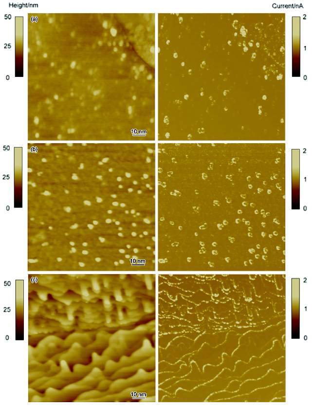
图1给出了用不同能量H离子辐照后钨样品的表面形貌图(左)和同时测得的电流分布图(右)。CAFM测量时在样品上施加负偏压就意味着电子流的方向是从样品表面到PtIr涂层的针尖表面, 而施加正偏压就意味着是从纳米针尖表面发射电子。考虑到施加负偏压可以避免导电针尖的氧化, 所以实验中均采用负偏压进行扫描。之前的分析结果也表明[12], 无论是采取正偏压还是负偏压扫描的方式都不会影响样品的电流分布图像。如图1所示, H离子辐照对钨样品表面产生了明显的损伤。经过20 eV H离子辐照后, 样品表面出现了不均匀的凸起颗粒, 这说明离子能量较低时, H离子与钨样品的作用仅局限于样品表面。在电流图像上也出现了与形貌图像相似的电流点分布。随着离子能量增加至120 eV, 样品表面出现了均匀的纳米尺寸凸起颗粒, 且这些颗粒的尺寸在50 nm左右。当能量增加至520 eV时, 样品表面出现了大量的更小尺寸(<10 nm)的纳米凸起物, 且这些凸起物呈现了具有一定方向性的条纹结构。这些条纹结构的形成可能与某个晶粒的晶面取向有关。电流分布图中的纳米尺寸缺陷点的大小和分布形态与同时测得的形貌图保持了高度的一致性, 这就说明这些表面凸起的产生可能与内表面缺陷的产生具有直接的关联性。Yang等[11]采用低能He离子辐照钨样品后也发现了类似的现象, 他们认为样片表面的凸起与氦泡的产生有关。由此, 认为低能H离子辐照后钨样品表面的纳米尺寸的凸起也可能是由于辐照后样品内部形成了纳米尺寸的氢泡缺陷, 当氢泡内聚集较多氢原子后, 氢泡逐渐长大, 并向样品表面迁移, 当样品内氢泡压力大于钨样品表面的屈服应力时, 氢泡内的超高压力就会向样品表面释放, 导致样品表面产生肿胀, 形成表面凸起。
图1 CAFM测得的不同能量的H离子辐照后钨样品的表面形貌(左图)和电流分布图像(右图)
Fig.1 Topography (left) and simultaneously measured current image (right) obtained using CAFM of H ions irradiated W samples with energy of 20 eV (a), 120 eV (b) and 520 eV (c)
为了考察低能H离子对高能离子预注入钨的辐照损伤作用, 将钨样品先经过高能(20 MeV) W6+室温注入后, 将钨样品暴露于低能H等离子体中, 进行辐照实验, 辐照温度为873 K, 剂量为1×1024 ionsm-2。采用CAFM分析了不同能量的H离子辐照预注入钨样品的表面形貌和同时得到的电流分布(图2)。如图所示, 随着辐照离子能量的增加, 样品表面纳米尺寸的凸起颗粒逐渐增加, 且均匀的分布于样品表面, 在不同实验条件下均没有观察到条纹状的表面微结构生成。电流分布图像中显示样品表面的缺陷点分布与形貌图像分布呈现高度的一致性, 而且随着辐照离子能量的增加, 导电缺陷点的大小和均匀性都在逐渐增加, 也没有出现条纹状分布的特征。与无预注入实验的钨样品相比, 经过预注入实验的钨样品表面形貌的凸起颗粒尺寸更小, 且分布更均匀。Tyburskad等[14]的研究表明, 高能离子预注入产生的离位损伤会大大增加氘的滞留量, 相对于无离位损伤的钨来说, 氘的滞留量将增加5-8倍。这是因为离位损伤产生的缺陷会吸附氘原子, 并成为氘原子的捕获阱而继续吸附氘原子, 导致氘原子滞留。因此, 20 MeV 高能W6+的预注入实验会在样品的注入区产生大量的空位、空位团簇、位错环等缺陷。这些缺陷的存在破坏了样品的有序性, 同时它们会成为低能离子辐照时捕获H原子的势阱, 不断吸引H原子聚集在这些缺陷周围, 从而降低H对钨样品表面的直接损伤作用。
图2 CAFM测得的不同能量的H离子辐照W6+预注入的钨样品的表面形貌(左图)和电流分布图像(右图)
Fig.2 Topography (left) and simultaneously measured current image (right) obtained using CAFM of H ions irradiated pre-implanted W samples with energy of 20 eV (a), 120 eV (b) and 520 eV (c)
图3给出了不同温度下H离子辐照无预注入钨样品的表面形貌和电流分布图像。如图所示, 辐照温度为673 K时, 钨样品表面出现了不均匀分布的凸起颗粒, 当温度增加至873 K时, 钨样品表面的凸起颗粒尺寸增加, 密度增加。这是因为温度的升高有利于纳米尺寸缺陷的迁移和聚集, 继而形成了较大尺寸的缺陷团。当温度增加至1073 K时, 钨样品表面出现了有方向性的条纹结构。从电流图像中也可以清晰地看到缺陷点的分布也呈现与形貌一致的方向性特征。
图3 CAFM测得的不同温度下H离子辐照后钨样品的表面形貌(左图)和电流分布图像(右图)
Fig.3 Topography (left) and simultaneously measured current image (right) obtained using CAFM of H ions irradiated W samples at 673 K (a), 873 K(b) and 1073 K (c)
图4给出了不同温度下H离子辐照高能离子预注入钨样品的表面形貌(左图)和电流分布图像。如图所示, 673 K时, 样品表面非常平整, 电流缺陷点的分布较少。当温度增加至873 K时, 样品表面出现了均匀分布的, 大小在20 nm左右的球状凸起颗粒(图2b)。当辐照温度增加至1073 K时, 样品表面也出现了具有一定方向性的条纹结构。这就说明当辐照温度升高至1073 K后, 高能离子预注入产生的缺陷会更容易复合。这些缺陷复合后将减弱H原子的吸附能力, 因此低能H离子与W样品的表面作用会更加显著, 导致样品的表面产生明显的损伤。
图4 CAFM测得的不同温度下H离子辐照W6+预注入的钨样品的表面形貌(左图)和电流分布图像(右图)
Fig.4 Topography (left) and simultaneously measured current image (right) obtained using CAFM of H ions irradiated pre-implanted W samples at 673 K (a) and 1073 K (b)
为了进一步分析钨样品表面的损伤特征, 对低能H离子辐照的钨样品和预注入的钨样品的表面形貌进行了SEM表征, 如图5所示, 在不同实验条件下辐照的钨样品表面均出现了严重的损伤结构。当辐照的离子能量≤120 eV或温度≤873 K时, 钨样品表面的损伤特征主要表现为球状或裂纹状表层结构(图5b), 在晶界的边缘出现表层即将脱落的迹象(图5a和d)。当温度增加至1073 K或离子能量增加至520 eV时, 钨样品表面出现条纹状的表层结构, 局部区域出现纳米丝状结构(图5c和e)。
图5 H 离子辐照后的钨样品表面形貌的SEM像
Fig.5 SEM images of surface of H ions irradiated W samples at 20 eV, 873 K (a), 120 eV, 873 K (b), 520 eV, 873 K (c), 120 eV, 673 K (d) and 120 eV, 1073 K (e)
图6给出了H 离子辐照W 6+预注入的钨样品表面形貌。从图中可以看出, 经过高能W 6+预注入的钨样品表面的损伤要明显低于无预注入的钨样品。在所有实验条件下, 样品表面出现的均为纳米尺寸的小颗粒, 这与CAFM的实验结果是一致的, 当辐照温度为1073 K时, 钨样品出现条纹状的表层结构, 但是并没有发现局部起丝现象。
图6 H 离子辐照W 6+预注入钨样品的表面形貌的SEM像
Fig.6 SEM images of surface of H ions irradiated W 6+ pre-implanted samples at 20 eV, 873 K (a), 120 eV, 873 K (b), 520 eV, 873 K (c), 120 eV, 673 K (d) and 120 eV, 1073 K (e)
图7给出了表面粗糙度随辐照离子能量和温度的变化曲线。如图所示, 钨样品的表面粗糙度均随着辐照能量和温度的增加而逐渐增加。有高能离子预注入的钨样品表面粗糙度要低于无预注入的钨样品, 进一步说明高能离子预注入实验可以降低低能H离子对钨样品的表面损伤作用。当温度增加至1073 K时, 高能离子预注入的钨样品表面粗糙度和无预注入的钨样品表面粗糙度相当, 这说明高温条件下, 高能离子预注入实验对低能H离子的辐照损伤作用开始减弱。
图7 表面粗糙度随辐照离子能量和温度的变化曲线
Fig.7 The influence of H ions energy (a) and temperature (b) on surface roughness
1.导电式原子力显微镜可以用于直接对比辐照后钨样品的表面凸起和纳米尺寸缺陷点分布之间的关系。分析表明, 辐照后钨样品表面的纳米尺寸的凸起是由于辐照后样品内部形成了氢泡, 为了释放内部压力向样品表面迁移, 导致表面肿胀的结果。
2. 低能H离子的能量和辐照温度变化对辐照后钨样品的表面微结构影响较大。 在H离子能量从20 eV到520 eV, 辐照温度低于1073 K时, 低能H离子对预注入样品的表面损伤作用要低于无预注入的钨样品, 这也就意味着高能离子预注入实验对钨材料的辐照损伤具有抑制作用; 但是当温度高于1073 K时, 这种抑制作用开始减弱。
The authors have declared that no competing interests exist.
| [1] |
Microstructure evolution in tungsten during low-energy helium ion irradiation ,
In situ transmission electron microscopy (TEM) study was performed to investigate the microstructural changes in tungsten during low-energy Heion irradiations in an electron microscope linked with an ion accelerator. The irradiations were carried out with 8 and 0.25 keV Heions at 293, 873 and 1073 K. In the case of the 8 keV irradiation, irradiation-induced vacancies act as nucleation sites for dislocation loops and helium (He) bubbles. Accordingly, such defects were formed even at the higher temperatures. With increasing irradiation temperature, the growth rates of dislocation loops and He bubbles rise remarkably. Although no vacancies are produced during 0.25 keV irradiation, He platelets, interstitial loops and He bubbles were formed. Impurity atoms may act as trapping centers for He atoms, which form bubbles by ejecting W atoms from their lattice site.
|
| [2] |
Ultra-fine grained/nano-crystalline tungsten-plasma facing materials for future fusion reactors ,超细晶/纳米晶钨-未来聚变堆面向等离子体材料 , |
| [3] |
Overview of the US-Japan collaborative investigation on hydrogen isotope retention in neutron-irradiated and ion-damaged tungsten ,
Plasma-facing components (PFCs) will be exposed to 14 MeV neutrons from deuterium-tritium (D-T) fusion reactions, and tungsten, a candidate PFC for the divertor in ITER, is expected to receive a neutron dose of 0.7 displacement per atom (dpa) by the end of operation in ITER. The effect of neutron-irradiation damage has been mainly simulated using high-energy ion bombardment. While this prior database of results is quite valuable for understanding the behavior of hydrogen isotopes in PFCs, it does not encompass the full range of effects that must be considered in a practical fusion environment due to short penetration depth, damage gradient, high damage rate, and high PKA energy spectrum of the ion bombardment. In addition, neutrons change the elemental composition via transmutations, and create a high radiation environment inside PFCs, which influence the behavior of hydrogen isotope in PFCs, suggesting the utilization of fission reactors is necessary for neutron irradiation. Therefore, the effort to correlate among high-energy ions, fission neutrons, and fusion neutrons is crucial for accurately estimating tritium retention under a neutron-irradiation environment. Under the framework of the US-Japan TITAN program, tungsten samples (99.99 at. % purity from A.L.M.T. Co.) were irradiated by neutron in the High Flux Isotope more» Reactor (HFIR), ORNL, at 50 and 300C to 0.025, 0.3, and 1.2 dpa, and the investigation of deuterium retention in neutron-irradiation was performed in the INL Tritium Plasma Experiment (TPE), the unique high-flux linear plasma facility that can handle tritium, beryllium and activated materials. This paper reports the recent results from the comparison of ion-damaged tungsten via various ion species (2.8 MeV Fe2+, 20 MeV W2+, and 700 keV H-) with that from neutron-irradiated tungsten to identify the similarities and differences among them. 芦less
|
| [4] |
The effect of displacement damage on deuterium retention in ITER-grade tungsten exposed to low-energy, high-flux pure and helium-seeded deuterium plasmas ,
ABSTRACT Samples prepared from polycrystalline ITER-grade tungsten were damaged by irradiation with 20MeVW ions at room temperature to a fluence of 1.4×10¹⁸W/m2. Due to the irradiation, displacement damage peaked near the end-of-range, 1.35μm beneath the surface, at 0.89 displacements per atom. The damaged as well as undamaged W samples were then exposed to low-energy, high-flux (1022D/m2s) pure D and helium-seeded D plasmas to an ion fluence of 3×1026D/m2 at various temperatures. Trapping of deuterium was examined by the D(3He,p)4He nuclear reaction at 3He energies varied from 0.69 to 4.0MeV allowing determination of the D concentration at depths up to 6μm. It has been found that (i) addition of 10% helium ions into the D plasma at exposure temperatures of 440–650K significantly reduces the D concentration at depths of 0.5–6μm compared to that for the pure plasma exposure; (ii) generation of the W-ion-induced displacement damage significantly increases the D concentration at depths up to 2μm (i.e., in the damage zone) under subsequent exposures to both pure D and D–He plasmas.
|
| [5] |
Formation of helium induced nanostructure ‘fuzz’ on various tungsten grades ,
The response of a variety of W material grades to nanostructure ‘fuzz’ formation is explored. W targets are exposed to He or D 2 –0.2He plasmas in PISCES-B at 900–132002K to below sputter threshold He + ions of energy 25–6002eV for up to 2.202×0210 4 02s. SEM and XPS reveal nanoscopic reorganization of the W surface to a layer of ‘fuzz’ of porosity 6590% as determined by a ‘fuzz’ removal/weight loss method. The variability of ‘fuzz’ growth is examined at 112002K for 102h durations: SR, SC and doped W grades – La 2 O 3 (1% wt.), Re (5% and 10% wt.), and TiC (1.5% wt.) developed 2–302μm thick ‘fuzz’ layers, while a VPS grade developed a layer 402μm thick. An RC grade revealed additional ‘fuzz’ at deep (>10002μm) grain boundaries. However, heat treatment up to 190002K produced reintegration of ‘fuzz’ with the bulk and He release at 65100002K and 651400–180002K due to depopulation from vacancy complexes.
|
| [6] |
Trapping of permeating deuterium in defect induced at the rear side of tungsten samples ,
As plasma-facing material in the Iter divertor area, tungsten will be subjected to bombardment with low-energy, high-flux deuterium and tritium particles, as well as 14 MeV neutrons. At elevated temperatures, hydrogen isotopes can diffuse into W material and can be trapped in n-induced defects. To account for this effect, the trapping of diffusing deuterium at defects generated in the rear side of the 25 and 500 mu m thick W samples damaged up to a depth of 2.2 mu m to a damage level of similar to 0.3 dpa was studied. The front side of each sample was exposed to a deuterium plasma at elevated temperatures. The results show that D atoms permeate through the whole width of the sample and are trapped in ion-induced defects. The highest D retention was measured at 460 K - beyond that point, due to the de-trapping process, it decreases with increasing irradiation temperature. (C) 2010 Elsevier B.V. All rights reserved.
|
| [7] |
Influence of helium-related defects on hydrogen retention in nano-polycrystalline tungsten films ,纳米多晶钨膜中He相关缺陷对H滞留的影响 ,
<p>用磁控溅射方法制备纳米多晶钨膜, 采用X射线衍射(XRD), 扫描电子显微镜(SEM), 弹性反冲探测(ERD)和慢正电子束分析(SPBA)等手段研究了在高能He<sup>+</sup>和H<sup>+</sup>依次对其辐照后He相关缺陷对H滞留的影响。结果表明, 注He<sup>+</sup>钨膜在退火后从<italic></italic>β型钨向<italic></italic>α型钨转变; 钨膜中的He含量随着退火温度的提高而减少, 在873 K退火加剧钨膜中He原子的释放, 且造成钨膜空位型缺陷的增加和结构无序度的提高; 钨膜中的H滞留总量随着He滞留总量的减少略有下降。</p>
|
| [8] |
Helium desorption in 3He implanted tungsten at low energy ,
The behavior of helium in He-3 implanted tungsten has been studied using Nuclear Reaction Analysis as a function of the post-implantation annealing temperature. Two different implantation conditions have been investigated: medium energy (60 keV), and low energy (0.3 keV), which exhibit drastically different helium release behavior. In the case of medium energy implantation, desorption starts from 1550 K and seems to be due to the dissociation of single helium-vacancy complexes (He-V-1). At 1873 K the released fraction reaches 75% that suggests the presence of a second type of helium trapping site. In the case of low energy implantation, desorption is observed from 400 K (slightly above room temperature) and indicates the presence of shallow helium traps the nature of which is discussed. The released fraction of helium saturates at similar to 60% at the temperature of 1473 K which could be due to helium trapping at single He-V-1 complexes. (C) 2010 Elsevier B.V. All rights reserved.
|
| [9] |
Blister formation on tungsten damaged by high energy particle irradiation ,
In order to investigate the effect of radiation damage on hydrogen behavior in tungsten, tungsten samples with radiation damage of up to 3.5 dpa were irradiated by a mixed hydrogen-carbon ion beam. The radiation damage was produced with 700 keV negative hydrogen ion beam irradiation. The number density of blisters produced by the mixed ion beam irradiation decreased with increasing radiation damage. This was especially observed for blisters with diameters of 20 渭m or less. This result showed that radiation damage produced by high-energy particle irradiation suppresses blister formation on tungsten surfaces.
|
| [10] |
Hydrogen behavior in damaged tungsten by high-energy ion irradiation ,
Effects of radiation damage on the behavior of hydrogen trapped in tungsten were investigated using a mixed hydrogen鈥揷arbon ion beam and deuterium ion beam. Radiation damage was produced by 300 and 700keV negative hydrogen ion beams. The number density of blisters formed on the radiation-damaged samples was less than an undamaged sample. SIMS measurements showed that post-implanted deuterium was mainly trapped at radiation damage sites. These results suggest that hydrogen accumulation at the grain boundaries is greatly decreased due to significant trapping of hydrogen isotopes at radiation damage sites.
|
| [11] |
Microscopic evolution of pre-damaged and undamaged tungsten exposed to low-energy and high-flux helium ions ,
High-energy (260 keV) He+ pre-damaged and undamaged polycrystalline tungsten samples were irradiated with low-energy (220 eV) and high-flux (锝1021 ions/m2 s) He+ at a sample temperature of 873 K to a fluence of 1.0 脳 1025 ions/m 2. Microscopic evolution of these samples was carried out using non-destructive conductive atomic force microscopy and a nanohardness test. Analysis indicates that a large number of nanometer-sized protuberances of irradiated tungsten samples results from over-high internal pressure of nanometer-sized helium bubbles. Ordered and nanostructured helium bubbles with the same diameter and average spacing can be formed due to the self-trapping and self-organizing of helium atoms in the tungsten materials. In the case of pre-damaged, low-energy He+ irradiation results in a random distribution of nanostructured helium bubbles, indicating that high-energy He+ implantation results in serious irradiation damage of tungsten materials, acting as nuclei for helium bubbles. ? 2014 Elsevier B.V. All rights reserved.
|
| [12] |
Structural and electrical evolution of He ion irradiated hydrocarbon films observed by conductive atomic force microscopy ,
Polymer-like hydrocarbon films are irradiated with 100keV He ion at the fluences of 1.0×10 15 –1.0×10 17 ions/cm 2 or at the irradiation temperature ranging from 25 to 600°C. Conductive atomic force microscopy (CAFM) has been used to evaluate the nanoscale electron conducting properties of these irradiated hydrocarbon films. Nanoscale and conducting defects have been formed in the hydrocarbon films irradiated at a relatively high ion fluence (1.0×10 17 ions/cm 2 ) or an elevated sample temperature. Analysis indicates that He ion irradiation results in the evolution of polymer-like hydrocarbon into a dense structure containing a large fraction of sp 2 carbon clusters. The sp 2 carbon clusters formed in irradiated hydrocarbon films can contribute to the formation of filament-like conducting channels with a relatively high local field-enhancing factor. Measurements indicate that the growth of nanoscale defects due to He ion irradiation can result in the surface swelling of irradiated hydrocarbon films at a relatively high ion fluences or elevated temperature.
|
| [13] |
Irradiation damage in silicon carbide based on a multi-mode scanning probe microscope ,基于多模式扫描探针显微镜技术分析碳化硅的辐照损伤 ,
本文在600 oC对6H-SiC进行了He+辐照实验,离子辐照能量为100 keV,剂量为5×10¹⁵ ions?cm?2、1×10¹⁶ions?cm?2、3×10¹⁶ ions?cm?2和8×10¹⁶ ions?cm?2。本文采用多模式扫描探针显微镜技术,包括轻敲模式原子力显微镜、纳米压痕/划痕和导电模式原子力显微镜技术对样品辐照前后的表面损伤进行了分析。结果表明,随辐照剂量的增加,样品表面粗糙度逐渐增加,表面硬度逐渐下降。导电模式原子力显微镜能清晰地观测到样品表面氦泡分布形态,进一步说明材料表面的肿胀是由材料内部高压氦泡产生的。
|
| [14] |
Deuterium retention in self-damaged tungsten ,
Surface and sub-surface morphology and deuterium retention in polycrystalline tungsten, undamaged and pre-damaged with 5.5 MeV W ions to damage levels of 0.04, 0.4, and 1.2 dpa, with and without subsequent annealing at 1200 K for 2 h, have been examined after irradiation with low-energy deuterium ions up to a fluence of 1 脳 10 D/m at various temperatures. The morphology changes were investigated by scanning electron microscopy in combination with focused ion beam surface cutting. The different hydrogen binding states were investigated by thermal desorption spectroscopy, and the depth profile of the implanted D was measured by ion beam nuclear reaction analysis. The dependence of the D retention on the damage level and the implanted fluence was studied, and the results show saturation for 0.4 dpa independent of the deuterium implantation fluence. Additionally, it was observed that self-implantation introduces high temperature traps which can almost completely be removed by annealing at 1200 K.
|

/
| 〈 |
|
〉 |


